Core Research Microscope Types
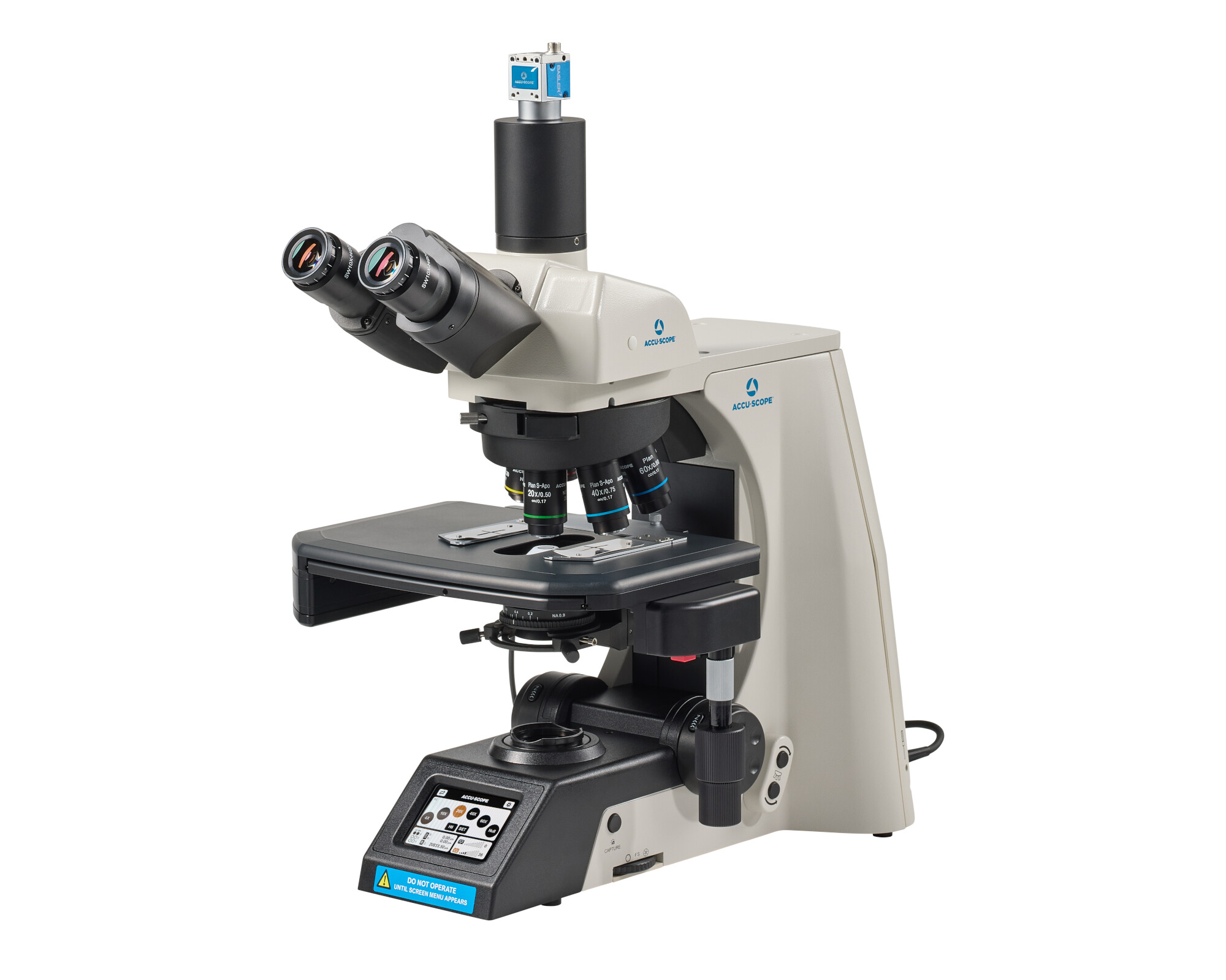
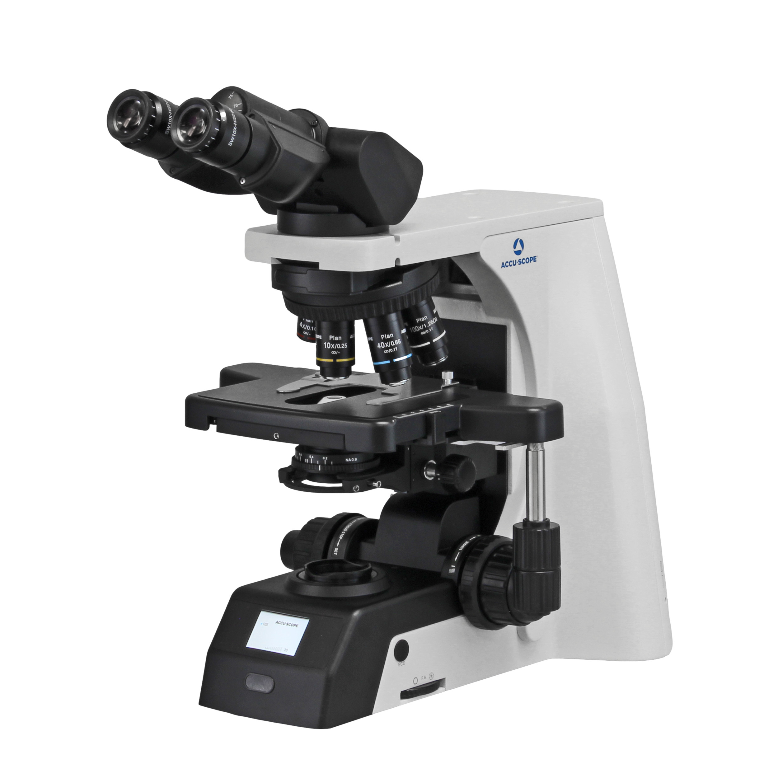
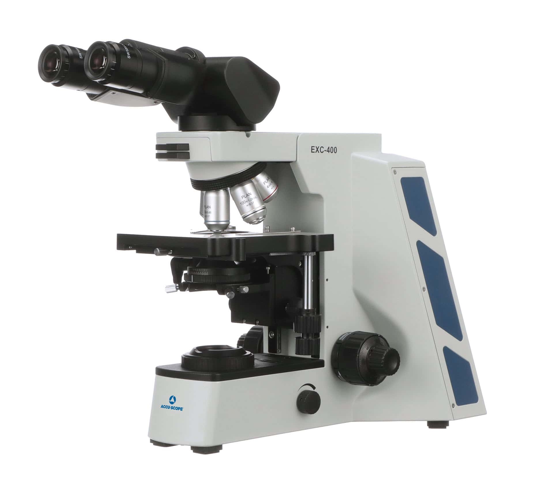
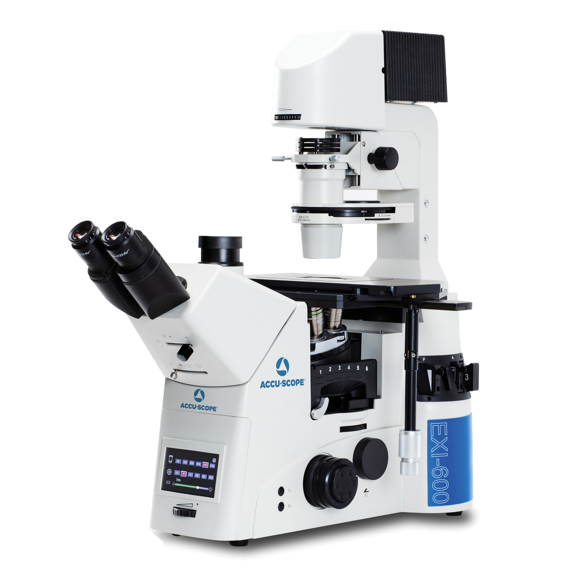
Compound microscopes use objective and eyepiece lens systems to produce the higher magnifications required for cellular and tissue imaging. Upright research microscopes are commonly used in histology, pathology, microbiology and cell biology to examine thin, prepared specimens on glass slides.
For routine histology and pathology, a configuration might include plan-corrected 4x, 10x, 20x and 40x objectives, with a 100x oil-immersion objective added when required. Highernumerical-aperture objectives collect more light and resolve finer detail, but should be evaluated alongside working distance and field flatness.
Stereo microscopes provide lower-magnification, three-dimensional views with generous working distance. These characteristics support tissue dissection, specimen sorting and manipulation, as well as inspection of opaque samples, small components and gems. Choose a zoom range that covers the required overview and detail, then confirm there is enough room beneath the objective for tools and sample handling.
For large or irregular samples, a boom stand can extend the microscope over the work area. A trinocular head provides a camera path for imaging and documentation. External illuminators can improve visibility on textured or reflective surfaces.