Posted on

ACCU-SCOPE Introduces Remote Collaboration Microscopy System for Pathology and Research

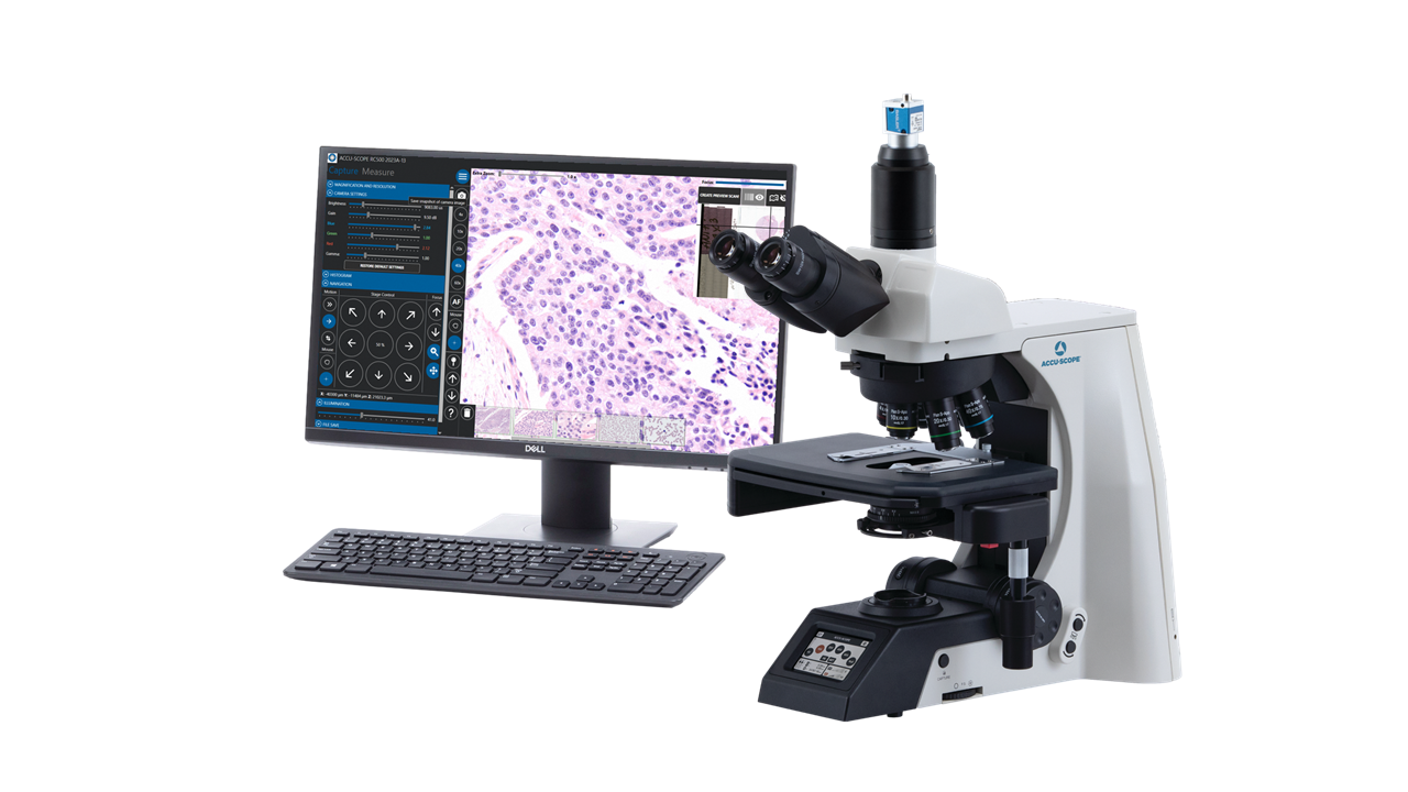
Commack, NY [June 2023] – ACCU-SCOPE Inc. is pleased to announce the development and launch of the RC500 Remote Collaboration Microscopy System.  The RC500 is fully integrated with motorized microscope, high-resolution, medical device-grade camera, and software.  The RC500 is designed for stand-alone operation for local, in-person microscopy, as well as full operation by a remote user via a desktop sharing application.  The RC500 provides the best of three worlds: traditional microscopy using eyepieces, improved ergonomics using the included camera and display on the monitor, and remote viewing of images by researchers and collaborators.

“Despite the movement of clinical microscopy towards whole slide imaging, there is still and absolutely a place for traditional microscopes in the clinic, laboratory, tumor boards, seminar and instructional environments,” says Brian Taub, CEO of ACCU-SCOPE.  “This has been a resounding message from pathologists, veterinary pathologists, and researchers over the past couple of years.  Slide scanners are indeed great, but they haven’t replaced the traditional microscope.”  The RC500 is a highly versatile system addressing the needs for peer-to-peer collaboration, remote consultation, and routine microscopy, and slide scanners don’t offer all three.  The RC500 delivers exceptional images plus the added benefit of excellent agility in today’s busy world of pathology and research.

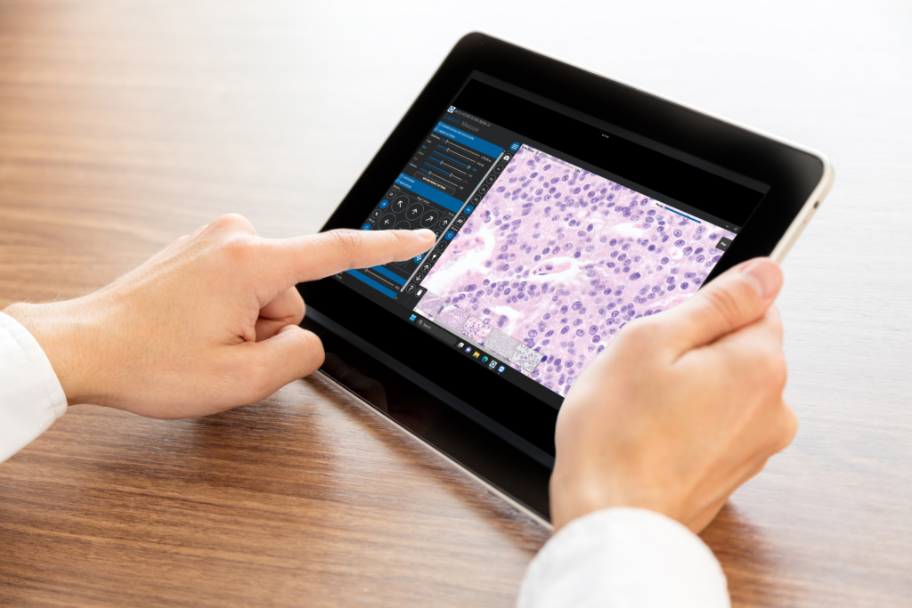
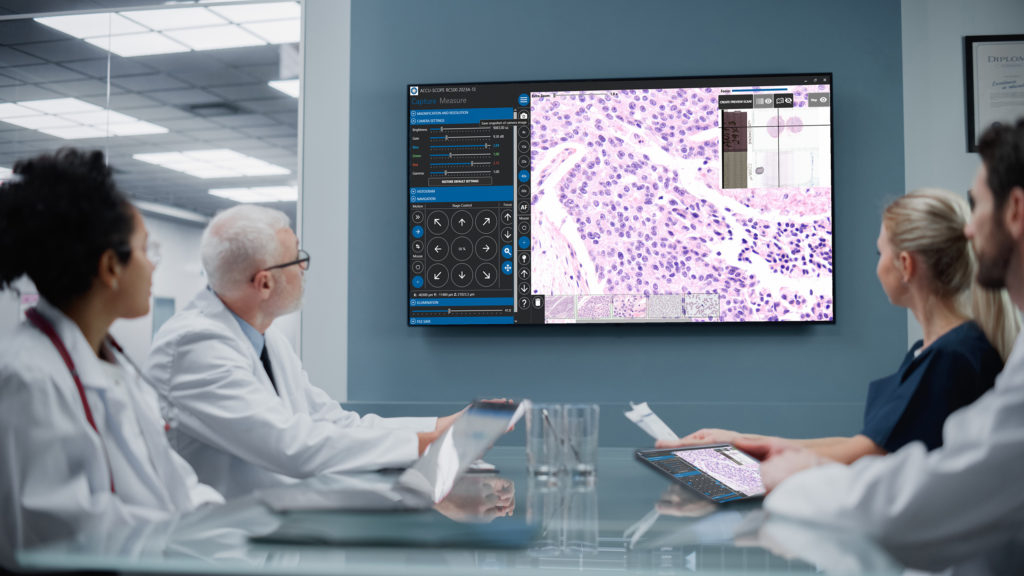
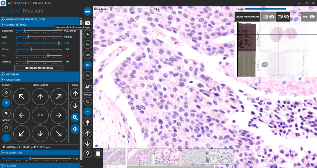
Figure 1. RC500 user interface.

Figure 2. RC500 system can be operated remotely from any PC, tablet or smartphone with internet connection and desktop sharing app.
Figure 3. The RC500 offers real-time remote viewing, control and collaboration. Remote viewers and users can position the slides, change objective magnifications and annotate for a true interactive experience.

The RC500 is a fully functional clinical and research-grade upright microscope featuring a trinocular viewing head; 4x, 10x, 20x, 40x and 60x dry semi-Apochromatically corrected objectives (no need for oil immersion); motorized stage with XY stage stalks and 2-slide holder; front touchscreen LCD display on the microscope; high-speed medical device-grade color microscopy camera; and preconfigured RC500 software.  RC500 provides both the local and remote user with fingertip access to objective selection, “manual” and auto focusing, XY specimen positioning, digital zoom, image adjustment, overview scan, pin drops, heat map, annotations, and more.

Contact ACCU-SCOPE for more information about the microscope or to schedule a remote demonstration of the RC500 Remote Collaboration System.

####

About ACCU-SCOPE Inc.

As a market leader in quality microscopy and imaging solutions, ACCU-SCOPE blends precision, performance and outstanding value like no other manufacturer can. We deliver superb yet affordable optical instrumentation and microscopy innovations that help advance the work, improve the outcomes and empower the achievements of the demanding professionals we’re privileged to serve. For more information about ACCU-SCOPE, please contact info@www.accu-scope.com.

Posted on

NEW Plan APO Objectives from ACCU-SCOPE

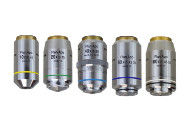
Commack, NY, USA [April 2023] – ACCU-SCOPE is pleased to introduce a line of Apochromatically-corrected (“APO”) objectives for upright (compound) and inverted microscopes.  The APO objectives complement the existing line of high-quality Plan Achromat and Plan Semi-Apochromatic objectives.

The new APO objectives were designed for the EXC-500 microscope – ACCU-SCOPE’s flagship clinical and biological upright microscope – the new APO lenses are also suitable for use on ACCU-SCOPE’s EXI-410 and EXI-600 inverted microscopes, provided the sample slides use a standard No. 1½ coverglass (0.17mm thickness).  Featuring higher Numerical Apertures and improved color correction than the Semi-Apochromatic or Achromat objectives, APO objectives deliver brighter images, shallower depth of field and higher resolution for improved observation of fine details.  APO objectives are ideal for brightfield observation, imaging and fluorescence applications, or where the highest color fidelity is desired.  The M25 thread size and 200mm tube length also allow these objectives to be used on most Nikon upright microscopes.

For more information, contact us at info@www.accu-scope.com.

####

Contact Us to learn more

Posted on

A Look at Stereo Microscopes and Their Applications

There is a wide array of microscopes available for scientists to use, each with their own functions, features, capabilities and applications. The stereo microscopes offer users a variety of unique benefits that aren’t provided by other microscopes. Here is an overview of this unique style of microscope, what makes it different, and why it has found application throughout countless industries.

What Is a Stereo Microscope?

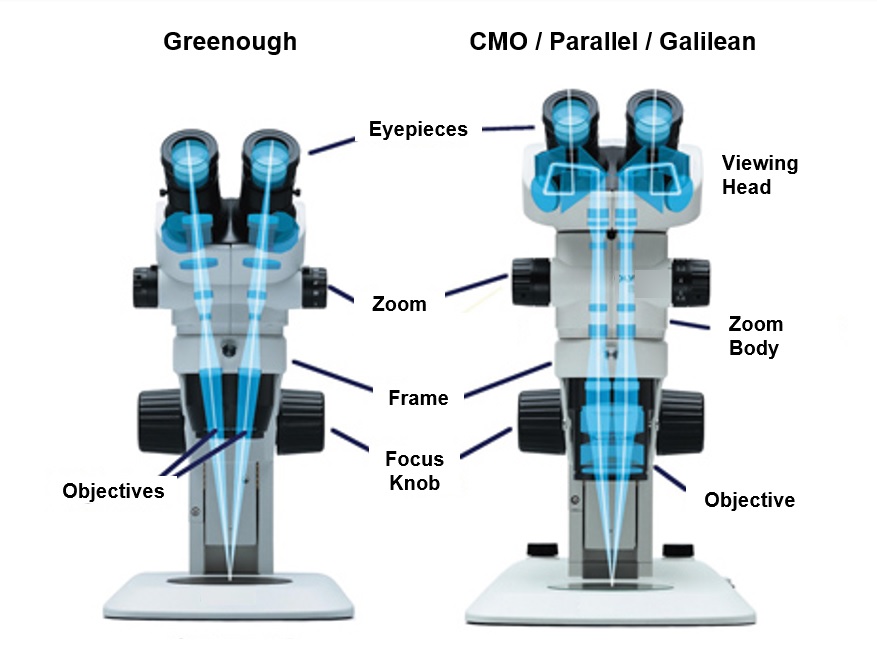
Stereo microscopes, also referred to as dissecting microscopes, examine 3-dimensional (3D) specimens, as opposed to conventional microscopes, which observe what are essentially 2-dimensional (flat) samples on slides. The objective lenses and eyepieces of stereo microscopes are independent, creating two distinct optical paths for the user’s eyes.  The optical paths begin at the eyes and converge at the focal position on the sample, thereby generating the 3D view (see the figure below). Zoom stereo microscopes allow adjustable magnification for more precise viewing, whereas some stereo microscopes may only have fixed magnifications. Auxiliary lenses are often available and allow the user to adjust the magnification to better fit their needs and/or the working distance (distance between the bottom of the microscope and the focal position on the sample).

Resulting from the optical design, stereo microscopes are fundamentally different from compound microscopes. The stand of a stereo microscope is almost as important as the microscope itself.  The stand can make a difference in how the sample is illuminated: from the top, bottom, on angle, or combinations of these, and the stand and illumination method are often selected based on the sample and the best way to observe it. Transmitted-style stands shine light from underneath and through the sample, which works best with transparent specimens. Incident lighting stands illuminate the sample from above or from the side using a light that is attached to the stand, a ring light, or other techniques. By controlling the angle of the incident light, small details can be revealed and more easily observed by the operator.

Additionally, stereo microscopes generally operate at lower magnifications than other types of microscopes and therefore generally, due to the physics of light and optics, have larger working distances (distance between the bottom of the microscope body and the top of the sample). In combination with the 3D viewing, the large working distances allow the operators more room for manipulation (e.g., dissection), the use of tools and the ability to reposition.  As a result, stereo microscopes.

Two Types of Stereo Microscopes?

That’s right, there are two distinct optical designs for stereo microscopes.  Stereo microscopes with a Greenough optical design have two completely independent optical paths.  The Greenough-design is what is typically associated with dissecting microscopes. Common Main Objective (CMO), Parallel or Galilean optical design stereo microscopes are distinguished by a large objective on the bottom of the microscope’s optical body.  CMO stereo microscopes generally provide higher magnification and higher resolution than their Greenough cousins.  As a result, CMO microscopes are more frequently used for imaging and documentation than dissection and sample manipulation – the exception is when observing specimens such as embryos, organoids and spheroids.

What Is a Stereo Microscope Used For?

The stereo microscope is superior to compound microscopes in viewing three-dimensional samples and their surface characteristics, making it the tool of choice for tasks requiring direct interaction with the material. As a result, they have many applications across a wide range of industries.  In academic settings, stereo microscopes are frequently used for dissections of both plant and animal samples, observation of model organisms (e.g., fruit flies, worms and fish) and other uses thanks to the zoom ranges, excellent depth perception and ease of specimen handling. Stereo microscopes are generally more portable than other microscopes, providing greater convenience and suitability for field work. Furthermore, the 3D viewing offered by stereo microscopes has led to their usage beyond biology dissections — botany, geology, gemology, and industrial applications capitalize on the unique capabilities of stereo microscopes for the observation of surface details and sample manipulation.

For High-Quality Stereo Microscopes and Other Products, Contact ACCU-SCOPE Today

If you would like to acquire high-quality stereo microscopes for your laboratory, reach out to ACCU-SCOPE. We offer various stereo microscope systems, microscope accessories, and a wide range of other products. To learn more about what a stereo microscope is best for your application, contact ACCU-SCOPE for additional information.

Posted on

See ACCU-SCOPE Microscopes at VMX 2023

ACCU-SCOPE Inc. is pleased to announce that several of its clinical microscopes are on display at VMX 2023 in Orlando, FL this week.  ACCU-SCOPE clinical microscopes are proven to deliver robust and dependable performance at a reasonable price.

The EXC-500 motorized microscope (EXC-500-MOT) is ACCU-SCOPE’s newest microscope and features motorized focus, a motorized nosepiece, an LCD touchscreen display and finger-tip hot buttons for fast changing of objectives and illumination intensity.  The EXC-500-MOT is key component in the Cellfie® Telemedicine Enabled Pathology System from Companion Animal Health (a division of LiteCure, LLC and DJO Global).  Cellfie® utilizes remote-enabled technologies to provide true point-of care pathology solutions for veterinary health.  See the Cellfie system in operation at the Companion Animal Health booth.

The ACCU-SCOPE 3000-LED and EXC-120 series microscopes are highly regarded by the veterinary community for their dependable operation and crisp images.  Each of these microscopes is on display at the Avante Animal Health booth this week at VMX 2023.  The 3000-LED is outfitted with an Excelis HD LITE camera with attachable HD monitor.  The Excelis HD LITE features built-in software for stand-alone operation without a PC.  The SKY WiFi 2 WiFi-enabled camera is shown on the EXC-120 microscope.  Simply download the SKYE View 2 mobile app to an Android or Apple mobile device, scan the QR code on the top of the SKYE WiFi camera and the specimen images are displayed right on your mobile device.  You can also capture and share images directly from your mobile device.  For more information or to see a demonstration, please visit Avante Animal Health in booth 1308.

####

As a market leader in quality microscopy and imaging solutions, ACCU-SCOPE blends precision, performance and outstanding value like no other manufacturer can. We deliver superb yet affordable optical instrumentation and microscopy innovations that help advance the work, improve the outcomes and empower the achievements of the demanding professionals we’re privileged to serve.

Posted on

Understanding Depth of Field and Depth of Focus in Microscopes

Depth of field and depth of focus are both well-known terms in photography, filmography, and microscopy. Given how similar their names are, they have sometimes been used interchangeably. However, despite both playing a vital role in camera and microscope optics, the two are very much different. Let’s look more closely at what defines the depth of field and depth of focus, their role in microscopy, and why the two should not be confused with one another.

Depth of Field

The depth of field is the distance between the closest object plane in focus and the furthest one in focus. In other words, it can be considered a lens’s ability to retain the quality of an image without moving the object farther or closer to the frame. Depth of field relates to how much detail can be seen above and below the optimal focus position.  When objects are placed at a set focal distance away from a lens, detail can blur and thereby degrade the resolution. As a result, smaller objects naturally lead to reduced depth of field for the lens. It is worth noting that the depth of field will increase as the condenser numerical aperture (condenser aperture diaphragm) is closed down (approaches zero).  While depth of field increases when the condenser aperture closes, resolution decreases.

Depth of Focus

Depth of focus is the imaging complement to depth of field and is more concerned with the tilt of the sensor plane of the microscope with respect to the lens image plane. This concept relates to how the quality of focus is altered as the object stays still and the lens’s sensor side changes. Therefore, the depth of focus will change significantly based on the magnification of the object and the microscope’s ability to focus light, known as the numerical aperture. Depth of focus has little to do with sensor size and is more closely related to sensor perpendicularity to the optical axis and pixel count — the more pixels the sensors have, the more noticeable sensor tilt will be on the quality of the image. Given how digital camera sensors are exposed at a given focal plane, depth of focus impacts the quality of the resulting image and is an essential consideration in photomicrography.

Depth of Focus vs. Depth of Field: How They Differ

As described above, depth of field and depth of focus relate to different perspectives. However, they are not entirely separate, as they both correspond to the axial depth of the space of a particular plane — depth of field relates to object space, whereas the depth of focus involves image space. This, in turn, influences their applications. For example, low-power objective lenses generally have lower numerical apertures and will have a higher depth of field (detail is lost quickly above or below the optimal focus plane) and higher depth of focus (sensor tilt is less impactful to image quality), while high-power objective lenses will have almost the reverse.

For Microscopes and More, Give Us a Call

Now that you have a better understanding of the depth of focus vs. depth of field differences and how they affect the quality of microscopes, see their influences for yourself with the help of ACCU-SCOPE’s high-quality microscope products. We offer a range of upright, inverted and dissection microscopes and various microscope accessories for laboratory settings and other environments. To learn more information about our products, give us a call today.

Posted on

ACCU-SCOPE Introduces ioLight Portable Digital Microscopes

Commack, NY – ACCU-SCOPE Inc. is please to introduce the line of ioLight portable digital microscopes to its portfolio.  Developed by ioLight Limited (UK), ioLight microscopes are designed for maximum portability and functionality in field work, the classroom and the laboratory.  The agreement with ioLight makes ACCU-SCOPE a master distributor of ioLight microscopes in North America, Latin America and the Caribbean, extending distribution through ACCU-SCOPE’s extensive dealer network.

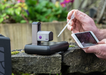
ioLight microscopes are currently available in 1mm and 2mm field of view (FOV) models, without or with a microscope slide XY stage to aide in slide positioning.  Equipped with top and bottom illumination, the user focuses on the specimen simply by sliding the camera head down the mast.  Fine focusing, illumination intensity and camera control are conducted from within the ioLight app on a smartphone or via a browser on any computer (Windows, MacOS or Chromebooks).  Snap an image or record a quick video and share it instantly via email or SMS.  The microscopes fold flat for storage and maximum transportability.

The size, convenience and performance of ioLight portable microscopes make them ideal for field studies where decisions can be made on the spot, eliminating the need to send samples to a lab and thereby saving scientists both time and money.  One very popular application is monitoring the nation’s waterways, lakes and oceans for harmful algal blooms (HABs).  HABs impact every state in the US and can cost the US economy tens of millions of dollars each year.  Field-based monitoring puts experts on location with the equipment they need to make rapid decisions and share their findings with colleagues and peers.  Read about NOAA’s $18.9M grant program for HAB research and monitoring: https://www.noaa.gov/news-release/noaa-awards-189m-for-harmful-algal-bloom-research-monitoring

Coming soon are improved and enhanced ioLight products including an updated inverted microscope (for observing samples in flasks or Petri dishes, such as cells in culture in incubators), and models of the 1mm, 2mm and inverted equipped for fluorescence microscopy and imaging. “CONTACT US for more information about ioLight portable digital microscopes.”

Posted on

How Microscopes Support In Vitro Fertilization

For centuries, biologists have been curious about the development of embryos. Because of the advancements in microscopy, our understanding of embryogenesis has not only evolved but now some of our best medical professionals use optical technologies to help women become pregnant.

When a woman and her partner struggle to conceive, they often turn to assisted reproductive technologies (ART) to achieve pregnancy. ART treatments include in vitro fertilization (IVF), intracytoplasmic sperm injection (ICSI), and intracytoplasmic morphologically-selected sperm injection (IMSI). Each method aims to fertilize a female’s egg outside of her body. For the procedure to go smoothly, the IVF laboratory must have high-quality microscopes to support ART.

Ways Microscopes Can Aid Assisted Reproduction

There are several reproductive medicine approaches that use different microscopy techniques to enhance a woman’s ability to become pregnant. These reproduction techniques are IVF, ICSI, and IMSI. Before fertilization can occur, an IVF scientist must take steps to prepare the sperm and egg. Each of these steps requires the use of microscopes. Let’s take a closer look at these procedures:

Semen Analysis

Before artificial fertilization can occur, an experienced scientist evaluates the quality of the sperm. They typically use an upright microscope to determine the total number, motility, and morphology of the sperm. Only healthy sperm will be used for fertilization. This pre-selection process requires a microscope with DIC or polarized light illumination.

Oocyte Preparation

After extracting oocytes from the female patient, the outer egg cell layers (besides the zona pellucida) are removed. This process is called “denuding” and is performed in a Petri dish while observing under a stereo microscope. The egg is transparent, so excellent illumination and contrast control are essential to be able to see the egg and its layers. Diascopic stands are the stereo microscope stands of choice and feature a tilting mirror to angle the light to provide oblique or darkfield contrast. Once this step is complete, the IVF scientist evaluates the morphology of the oocyte using an inverted microscope for its high magnification and easy accessibility to the egg for micromanipulation. If abnormalities are detected, the oocyte will not be used during fertilization.

Fertilization

Once the healthy sperms and oocytes have been isolated, artificial fertilization follows next. With ICSI, a single sperm cell is injected into an oocyte using a micromanipulator. The oocyte’s zona pellucida and polar body must be visible for this procedure. An alternative to ICSI is IMSI, which involves the extra steps of assessing sperm morphology using an inverted microscope and injecting sperm with the desired morphology.

Embryo Growth

After the eggs are fertilized, an embryologist monitors the growth and development of the embryos over the next few days. They look for any imperfections in the embryos. Only the best and healthiest embryos are implanted into the mother.

How to Choose a Microscope for IVF Applications

ART laboratories require a range of microscopes to complete various clinical procedures, such as gamete selection and embryo monitoring. Choosing the right microscope requires considering several factors, including the specific operation or application and the microscope’s features. When selecting a microscope for IVF applications, look for the following features:

Stereo Microscope

  • A zoom range that provides enough magnification to see detail in the egg
  • Diascopic stand that generates the necessary contrast to reveal the detail in the egg

Inverted Microscope

  • An inverted design that allows access to the cells or embryos for manipulation
  • A range of objectives to ensure sufficient magnification that facilitates observation of the specimen and the use of micromanipulation tools during sperm and egg cell handling
  • A built-in camera port to support the use of a digital camera for viewing specimens and documentation

Need Microscopes for IVF Procedures? Turn to ACCU-SCOPE

IVF laboratories need high-quality microscopes they can rely on. If you’re searching for stereo, inverted, and upright microscopes to outfit your IVF and embryology laboratory, you’ve come to the right place.

At ACCU-SCOPE, we have a wide selection of microscopes used for embryology and IVF, including stereo and inverted microscopes with cameras. We can even equip your lab with stereo stands and accessories that work with various microscope models.

Contact our team today for help choosing the right microscope for your application!

Posted on

The Best Microscopes for Educational Environments

An educator’s job is never done. Whether it’s the start or end of a school year, teachers and professors are constantly on the lookout for top-of-the-line microscopes for education. They want to equip their students with the tools they need to explore the unseen world around them, stimulate curiosity, and make incredible discoveries. Unfortunately, microscope shopping takes a great deal of time, and if they look in the wrong place, all their efforts could be for naught.

Fortunately, ACCU-SCOPE offers a wide selection of the best microscopes for educational environments. With ACCU-SCOPE, you will be able to outfit your classroom or school laboratory with the affordable and quality tools your students need — whether they’re high school students getting a first glimpse at biology or graduate school students conducting their own experiments.

The Top Microscopes for Middle School, High School, and College Students

As you shop for the classroom, you’ll recognize a variety of factors to consider before buying a student microscope. For example, you need to decide whether monocular or binocular microscopes are ideal for your students’ needs and which magnification levels will suit your application. In addition, you’ll need to consider the device’s frame, optics, lighting, field of view, and resolution. There’s a lot to think about!

ACCU-SCOPE created this easily digestible guide to help you buy the right microscopes for your classroom. Here are some of the best options for educational settings:

EXM-150 Microscopes

Middle school and high school students are still in the introductory phase to microscopes and science as a whole. This means they generally don’t require the advanced features or super high magnifications that may be more of a distraction than they are helpful. Of course, this doesn’t mean educators should compromise on quality. Great images and extreme durability are still important. That’s where the EXM-150 monocular microscope comes in.

The EXM-150 series provides great performance at an affordable price. Its student-proof design makes it the perfect instrument for any setting. Inexperienced students can make precise observations without having to fuss with various settings. The mechanical stage is highly secure, so you can depend on it for years to come. Even better, this microscope features LED lights that budding scientists can use with or without a cord, plus an integrated carrying handle, making the instrument easily transportable.

EXS-210 Stereo Microscopes

The EXS-210 series holds true to the adage, “Big things come in small packages.” This compact stereo microscope provides quality, versatility, and performance at an affordable price. It is student-proof, has corded or cordless operation, and is easily transportable, making classroom set-up a breeze. It also uses our state-of-the-art optical coating techniques and LED illuminators for high-quality images, durability, and dependability. This microscope is highly favored by middle and high school students, homeschoolers, and hobbyists.

EXC-120 Microscopes

If your classroom requires microscopes that can withstand years of rigorous use and you want binocular or trinocular viewing for greater comfort and image quality, you’ll want to consider the EXC-120 series. These microscopes are specifically engineered for clinical and veterinary applications, and classroom laboratories, making them ideal for college and graduate students. An EXC-120 microscope features high-contrast objectives, a die-cast frame, ergonomic focusing controls, brass gears, and a wide field of view. With this microscope, it’s never been easier to achieve clear images while observing your specimen in a comfortable position.

3079 Stereo Microscopes (Dissecting Microscopes)

If your students are ready to take their observations to the next level, the 3078 stereo microscope series is the best option for you. Its top-of-the-line optical system provides high resolution, 3-dimensional views of samples, while its ergonomic and durable design makes it ideal for long-term use. These microscopes are frequently found in university laboratories and industrial and OEM applications.

Going Digital: The Future of Microscopy in the Classroom

Whether you’re buying microscopes for high school or college labs, you will want to consider equipping the classroom or laboratory with digital microscopes. These microscopes either have a camera already installed or have connections that allow for the attachment of an LCD camera. With these modern instruments, you can demonstrate important scientific concepts in real time by projecting the image on a screen for all to see. Your students can also easily share their findings by saving the images or videos for a report.

We recommend the following digital cameras for educational purposes:

  • Excelis HD Lite Camera: This camera works with most microscopes and allows HD live imaging of samples. The built-in software allows streamlined operation without a computer. The user can also capture images using CaptaVision+ software for editing and measuring at a later time.
  • SKYE WiFi 3 Camera: This camera creates its own WiFi network so the user can easily stream live images to a range of connected Android and iOS devices. Simply download the SKYE View 2 app, scan the QR code on the camera, and you’re ready to go! Thirteen students or more can connect to a single camera and view images simultaneously.
  • Excelis 4K Camera: This camera delivers high-definition color images to any 4K monitor and the built-in software allows operation without a PC. Alternatively, the user can connect the camera to a PC using a USB 3.0 cable and control the camera through the CaptaVision+ imaging software.

Get the Best Microscope for Your Classroom Today

No matter your education needs, ACCU-SCOPE has a microscope that is perfect for you! We have an expansive range of educational microscopes plus the expertise to help you decide which ones will meet your students’ needs best. We continue to expand our product line to develop and include the latest advancements in the industry. Contact us today to learn more about our educational microscopy solutions and keep an eye out for new product introductions.
Posted on

Top Considerations When Buying A Microscopy Camera, PART 8: Controlling Your Camera (software)

Something that many people don’t consider enough is how the camera is controlled.  Whether by mobile app, built-in software or computer software, this user interface is the way you communicate with the camera, view your samples and capture images or video.

Computer-based software offer the highest level of control for the camera.  From acquisition settings to image processing, calibration/measurement and reporting, computer software provides maximum flexibility and features.  In most cases, the software is free.  The caveat is that the software may not perform exactly as you like.

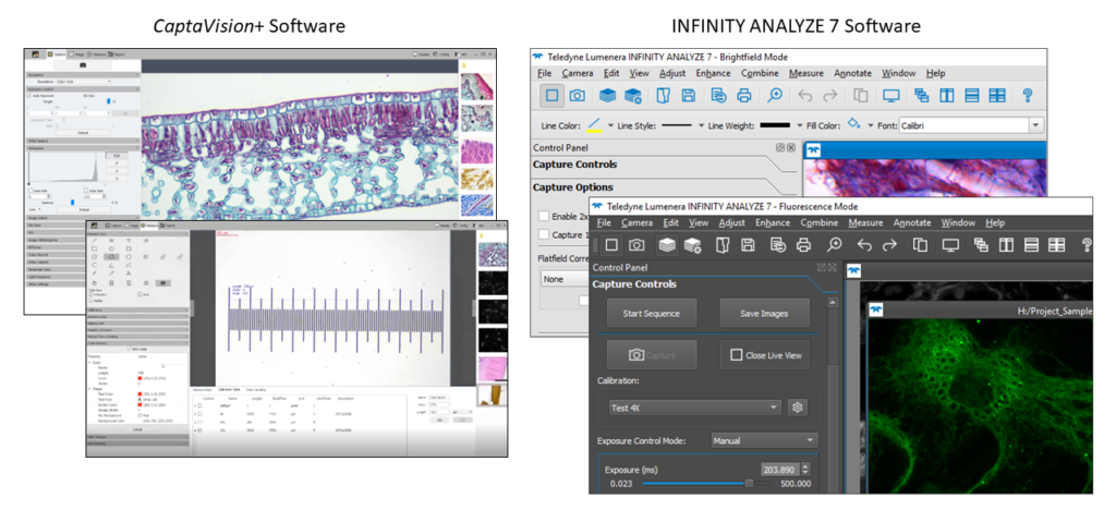
Figure 16. Examples of the graphical user interface for two software available from ACCU-SCOPE.  CaptaVision+ is a free software that is available with all ACCU-SCOPE Excelis camera.  INFINITY ANALYZE 7 is a free software that comes with all Teledyne Lumenera Infinity cameras.  The software are compatible only with the cameras specified.

One alternative may be to use software from a third-party supplier.  If the software doesn’t fully support the camera, it may be able to use a TWAIN driver to provide basic functionality and control of the camera.  Third-party software often includes the ability to control illumination sources, filter wheels, stages and even the microscope itself.  But all of this comes at a cost, as third-party software can be very specialized and expensive.  However, it can be worth the investment if you get the control and capabilities you need for your work.

More cameras are available with built-in software with basic acquisition controls and sometimes measurement functionality.  Usually controlled via a mouse, cameras with built-in software are ideal for stand-alone operation where there isn’t room for a PC or where the application only requires a minimal set of features.  These microscope and camera systems are generally more portable, too.

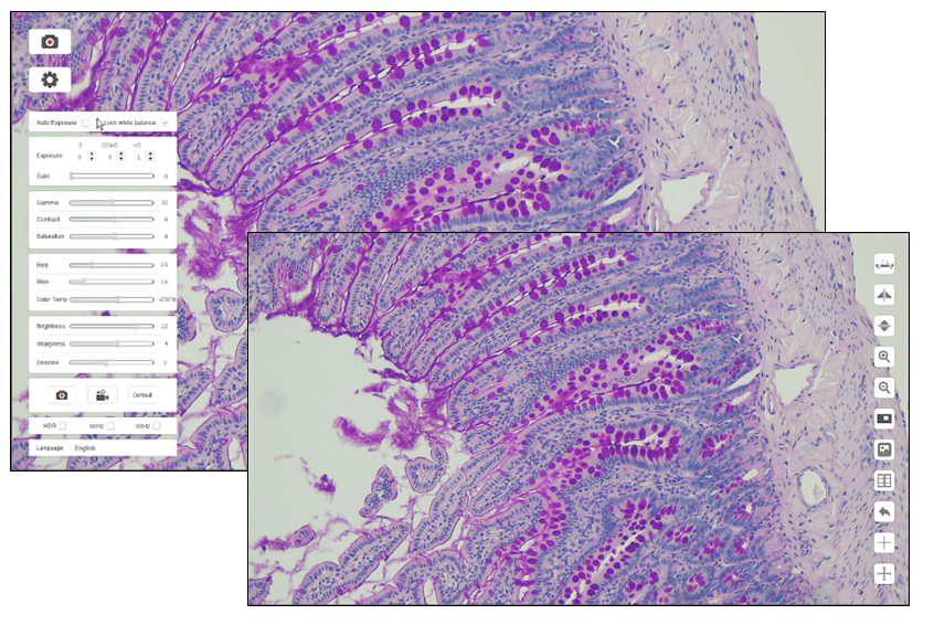
Figure 17. Graphical user interface of Excelis HD built-in software.  Notice the simplified settings menu (main image) and the measurement and annotation tools (inset image).

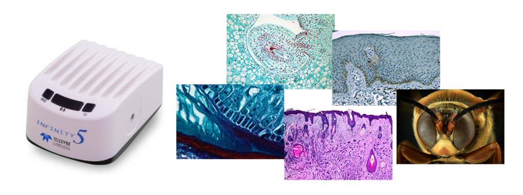
Some cameras have some level of built-in software, and the functionality has been reduced to a few buttons on the outside of the camera housing.  The Teledyne Lumenera Infinity 5 series are a great example of this, and they possess buttons only for power, white balance, and snapping an image.

Figure 18. Photo of the Teledyne Lumenera Infinity 5 series camera showing the easily accessible white balance, capture and power buttons.  On the right are sample specimen images captured with Teledyne Lumenera Infinity cameras.

Even cameras can now generate their own WiFi signals that are readily identified by our mobile devices.  Mobile apps offer the convenience of portability and multiple users, with some cameras supporting up to 10 or more simultaneously connected users.

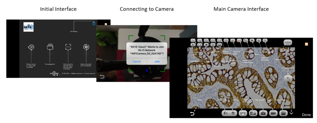
Figure 19. Screenshots of SKYE View 2 mobile app on iPhone. Initial Interface is seen when the app is started. Click the [ ] icon to enter the camera pairing mode. Point the mobile phone camera towards the QR code on top of the SKYE WiFi 2 microscopy camera, then click Join. Main Camera Interface illustrates the variety of camera and image processing controls.

Choosing a camera that suits your needs is one decision, but don’t ignore the camera control software.  Both are required to get the job done.

This blog post concludes our series “Top Considerations When Buying A Microscopy Camera.”  We hope you found it interesting and learned something about microscopy cameras.  If there is another topic relating to microscopy cameras that you would like us to discuss, please share it in the Comments section.

Posted on

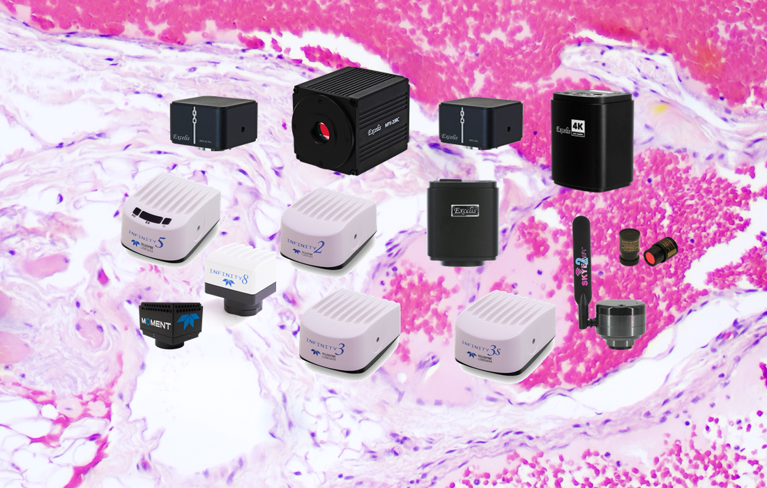
ACCU-SCOPE Confirms NDAA Compliance of Imaging Devices

Commack, NY, USA [August 2022] – ACCU-SCOPE is pleased to announce that all our microscopy cameras and imaging systems are fully compliant with Section 889 of the John S. McCain National Defense Authorization Act of 2019 (Sec.889, NDAA 2019) and do not contain any parts or components from prohibited companies as outlined in the NDAA.

NDAA Summary

The John S. McCain National Defense Authorization Act for Fiscal Year 2019 was signed into law on August 13, 2018. The law, specifically Section 889, amongst other things prohibits federal agencies, their contractors, and grant or loan recipients from procuring or using “telecommunications and video surveillance equipment or services” from specific prohibited companies as a “substantial or essential component of any system, or as critical technology as part of any system.”  The prohibited companies include Huawei Technologies Company or ZTE Corporation, Hytera Communications Corporation, Hangzhou Hikvision Digital Technology Company, and Dahua Technology Company; and their affiliated entities.

Catalog number Description

AU-300-EP

(discontinued) ACCU-CAM 300 3MP eyepiece camera

AU-500-EP

ACCU-CAM 500 5MP eyepiece camera

AU-300-HD

Excelis HD Lite color microscopy camera

AU-300-HDS

Excelis HD Lite color microscopy camera, with 11.6” monitor

AU-600-HD

Excelis HD color microscopy camera

AU-600-HDS

Excelis HD color microscopy camera, with 11.6” monitor

AU-800-4K

Excelis 4K color microscopy camera

AU-EC50

EC50 5.1MP color microscopy camera

AU-5C-CMOS

MPX-5C Pro 5MP color microscopy camera

AU-6C-CMOS

MPX-6C 6MP color microscopy camera

AU-20C-CMOS

MPX-20C 20MP color microscopy camera

AU-20RC-CMOS

MPX-20RC 20MP color microscopy camera, cooled

AU-200-HD

(discontinued) INFINITYHD color HD microscopy camera

AU-200-CMOS

INFINITY1-2CB 2MP color microscopy camera

AU-310-CMOS

INFINITY1-3C 3.1MP color microscopy camera

AU-500-CMOS

INFINITY1-5C 5MP color microscopy camera

AU-140RC-CCD

INFINITY2-1RC 1.4MP color microscopy camera

AU-140RM-CCD

INFINITY2-1RM 1.4MP monochrome microscopy camera

AU-200-CCD

INFINITY2-2C 2MP color microscopy camera

AU-200M-CCD

INFINITY2-2M 2MP monochrome microscopy camera

AU-330-CCD

INFINITY2-3C 3.3MP color microscopy camera

AU-500-CCD

INFINITY2-5C 5MP color microscopy camera

AU-140CL-CCD

INFINITY3-1C 1.4MP color microscopy camera, cooled

AU-140CLM-CCD

INFINITY3-1M 1.4MP monochrome microscopy camera, cooled

AU-140URC-CCD

INFINITY3-1URC 1.4MP color microscopy camera

AU-140URM-CCD

INFINITY3-1URM 1.4MP monochrome microscopy camera

AU-141SURC-CCD

INFINITY3S-1URC 1.4MP color microscopy camera

AU-141SURM-CCD

INFINITY3S-1URM 1.4MP monochrome microscopy camera

AU-280C-CCD

INFINITY3-3URC 2.8MP color microscopy camera

AU-280M-CCD

INFINITY3-3URM 2.8MP monochrome microscopy camera

AU-600URC-CCD

INFINITY3-6URC 6MP color microscopy camera

AU-600URM-CCD

INFINITY3-6URM 6MP monochrome microscopy camera

AU-320C-CMOS

INFINITY5-3C 3.2MP color microscopy camera with HDMI out

AU-320M-CMOS

INFINITY5-3M 3.2MP monochrome microscopy camera with HDMI out

AU-510C-CMOS

INFINITY5-5C 5.1MP color microscopy camera with HDMI out

AU-510M-CMOS

INFINITY5-5M 5.1MP monochrome microscopy camera with HDMI out

I8-LM00-02M

INFINITY i8 2MP monochrome microscopy camera

I8-LC00-08M

INFINITY i8 8MP color microscopy camera

I8-LM00-08M

INFINITY i8 8MP monochrome microscopy camera

I8-LC00-20M

INFINITY i8 20MP color microscopy camera

AT-MOMENT-MONO

Moment Monochrome microscopy camera

About ACCU-SCOPE Inc. 

As a market leader in quality microscopy and imaging solutions, ACCU-SCOPE blends precision, performance and outstanding value like no other manufacturer can. We deliver superb yet affordable optical instrumentation and microscopy innovations that help advance the work, improve the outcomes and empower the achievements of the demanding professionals we’re privileged to serve. For more information, please contact info@www.accu-scope.com.