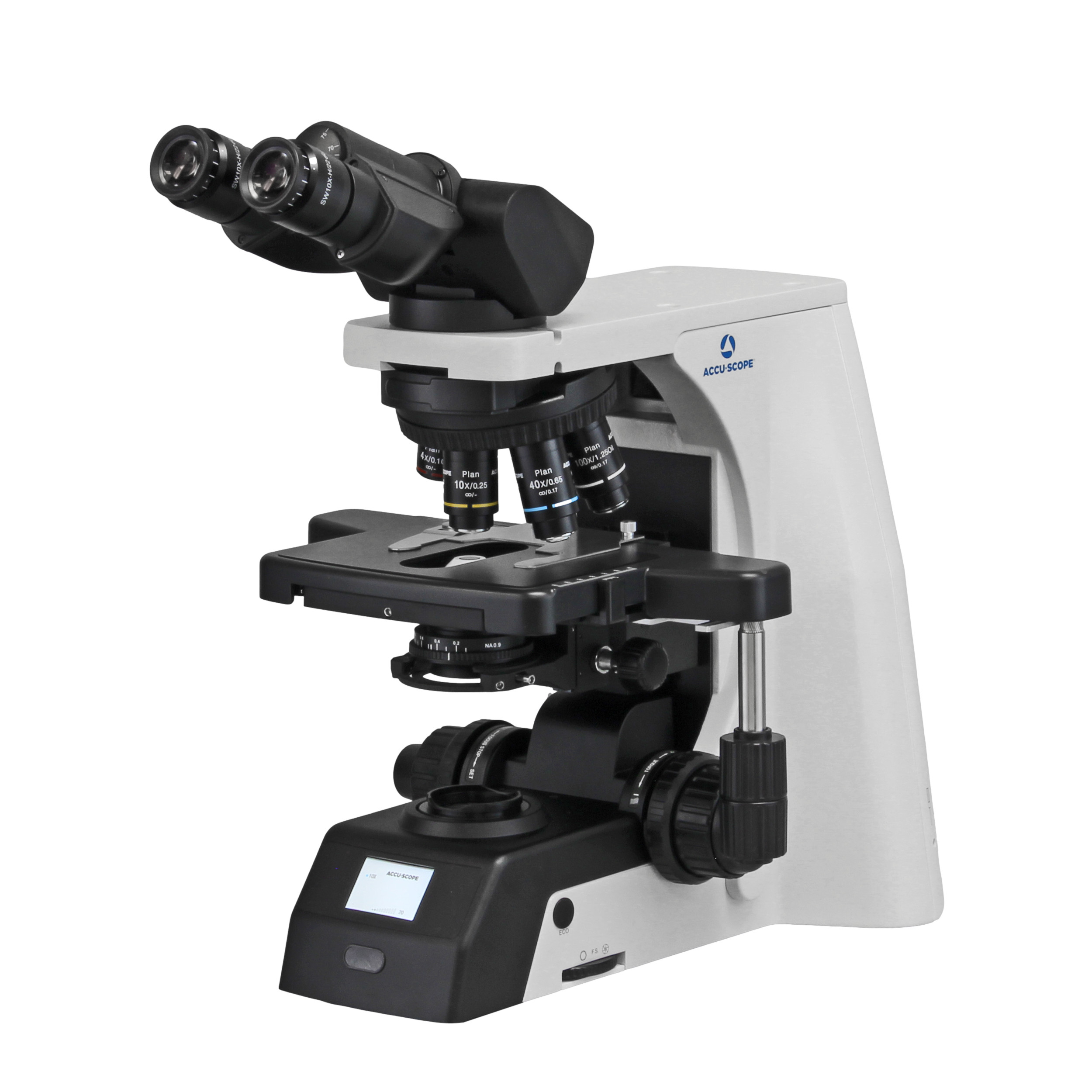
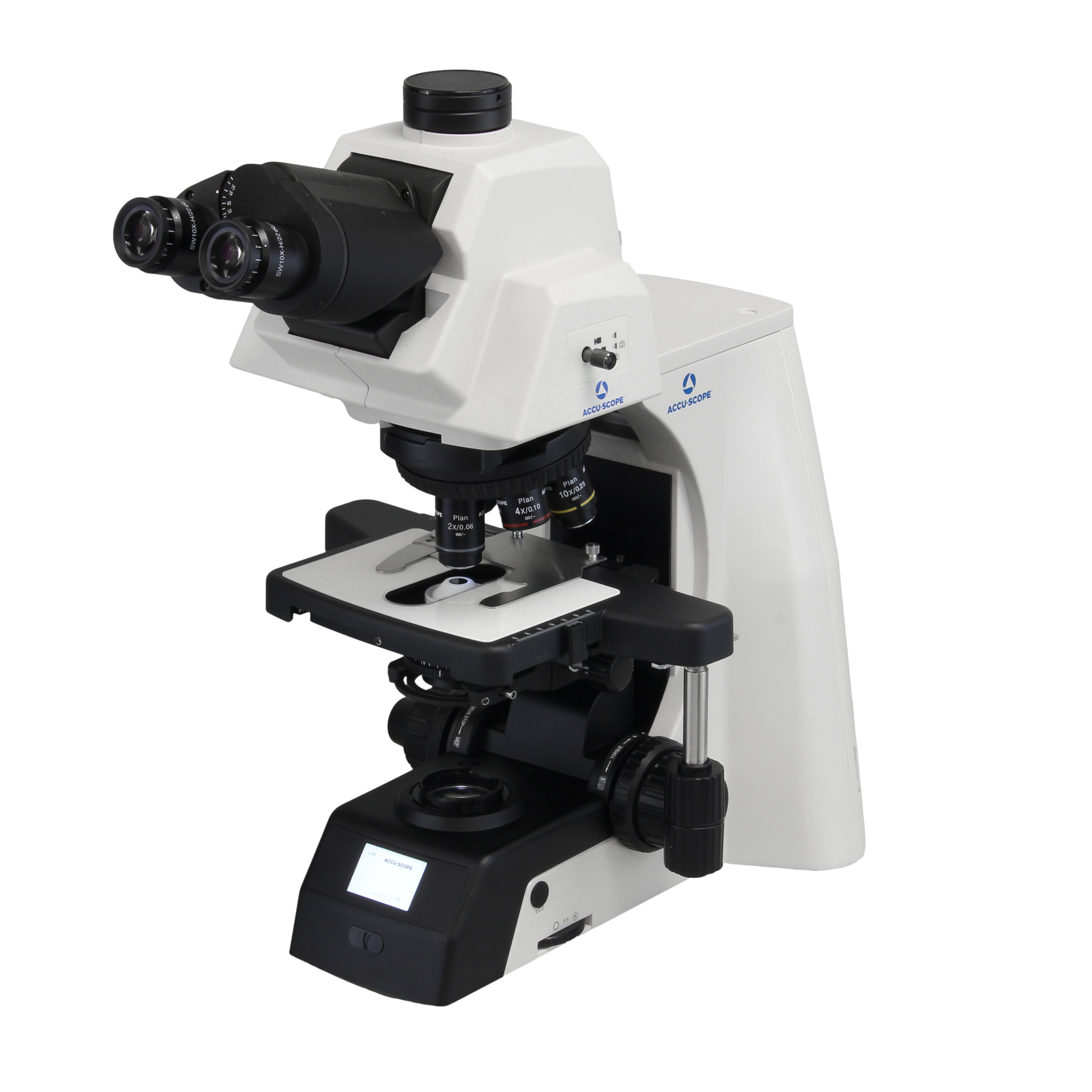

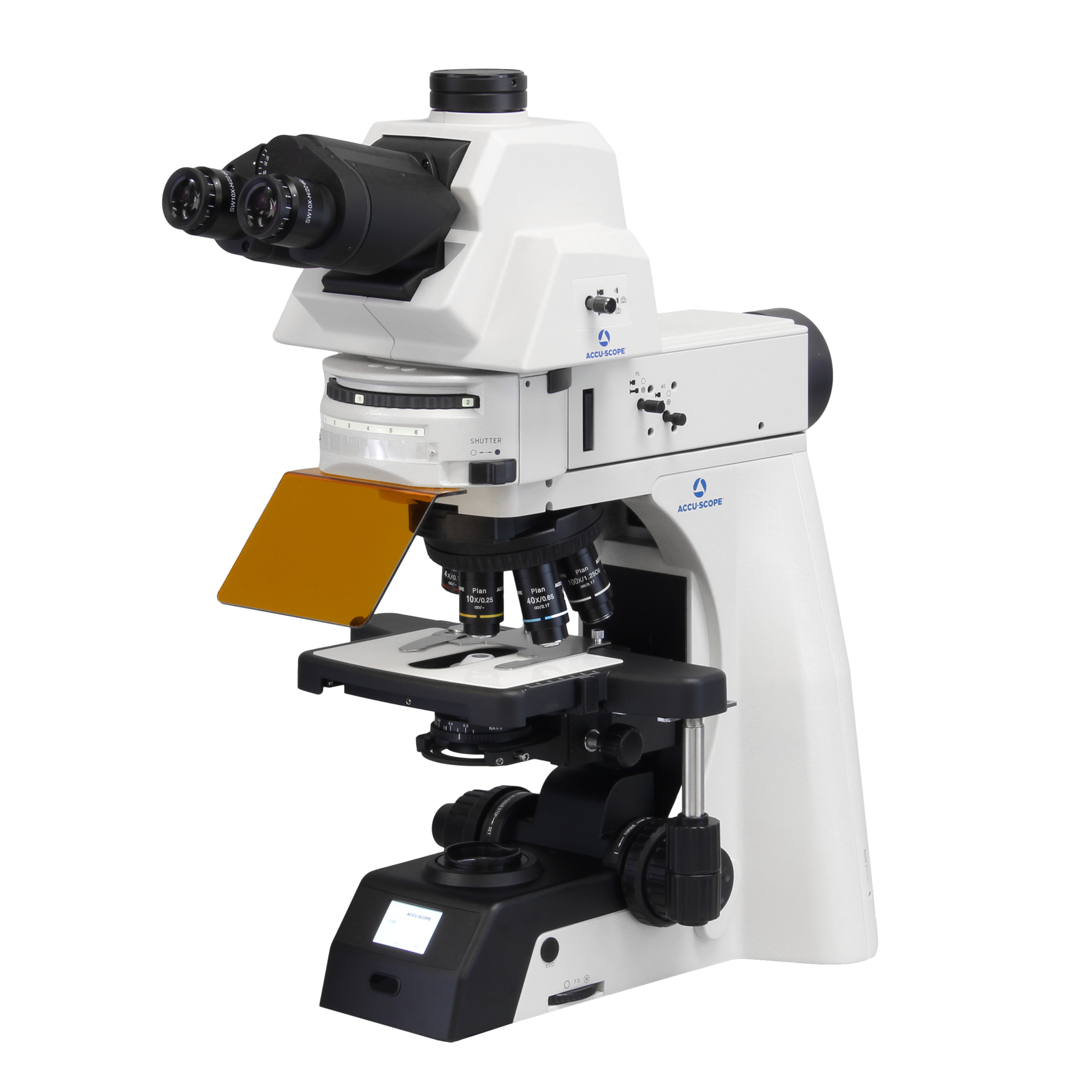


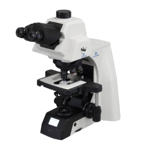
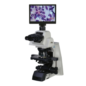
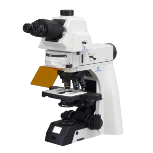
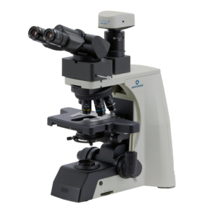
ACCU-SCOPE’s flagship EXC-500 Microscope offers best-in-class performance and value for clinical laboratory and research applications. The NIS infinity optical system provides sharp, crisp images with outstanding detail. Its modular design allows the EXC-500 to accommodate a vast array of research, fluorescence, phase contrast, DIC and darkfield applications. The EXC-500 features optimized LED-based ECO-illumination capable of reproducing the sensitive color differences often seen in many clinical samples. Add one of our many digital imaging solutions for image capture, image analysis, documentation and sharing.
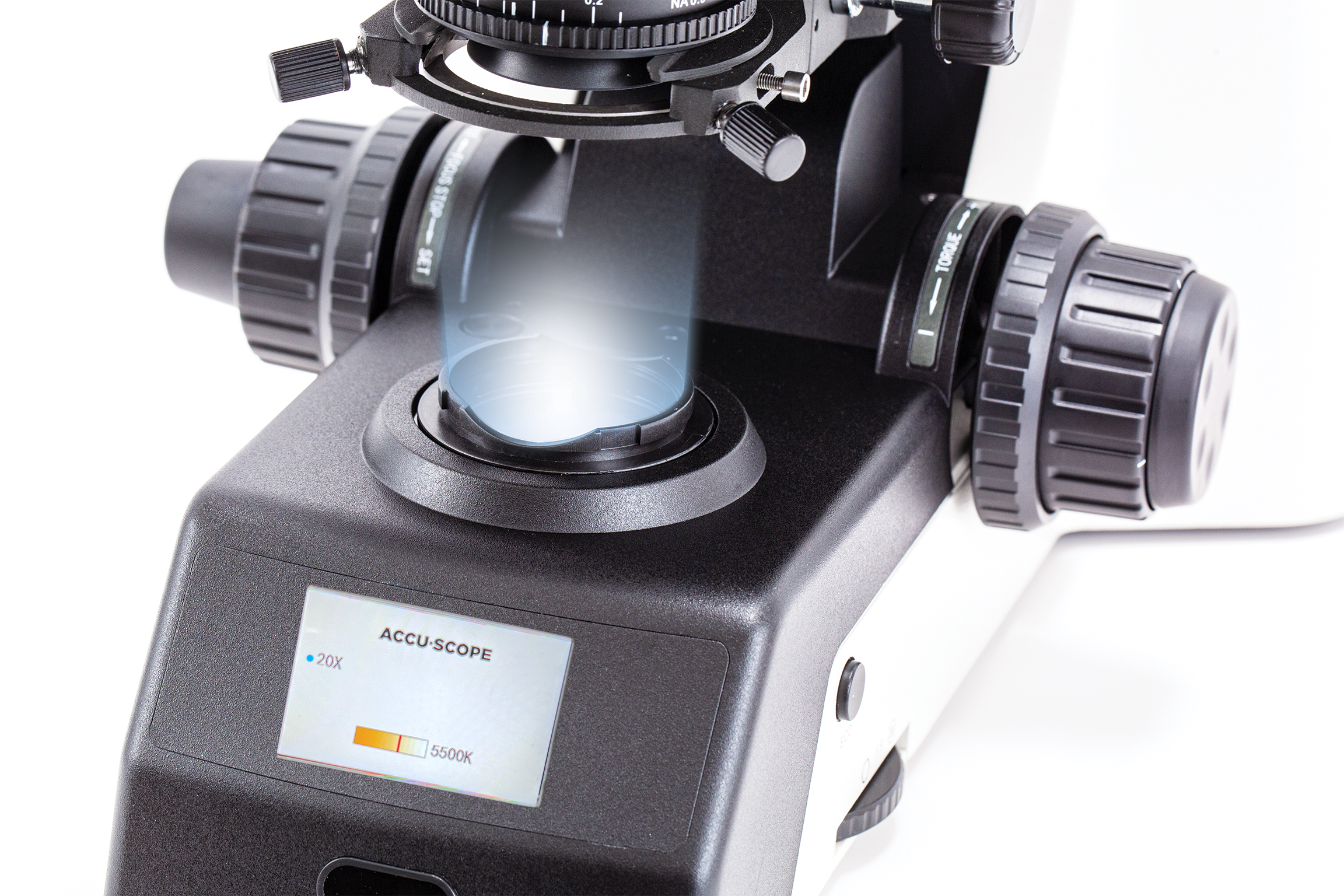
The new programmable LCD screen enables a personalized experience with the EXC-500. The user can set light intensity and color temperature by objective. The LCD displays the current objective status, and the user can also set Sleep timer preferences, the LCD background color, and lock the settings with a password.
Engineered for pathology and cytology, the EXC-500 uses a white LED with a high color rendering index allowing users to visualize samples in true color. The high-powered white LED allows for constant color temperature and fast, efficient operation for digital documentation.
The new EXC-500 features Light Intensity Memory and Color Temperature Memory, now allowing the user to set color temperature and light intensity by objective, for a truly personalized observation experience.
The EXC-500 is designed for maximum ergonomic efficiency to allow for long periods of use. Each of the primary controls can be adjusted with one hand – focus, tension adjustable stage controls, and field diaphragm.
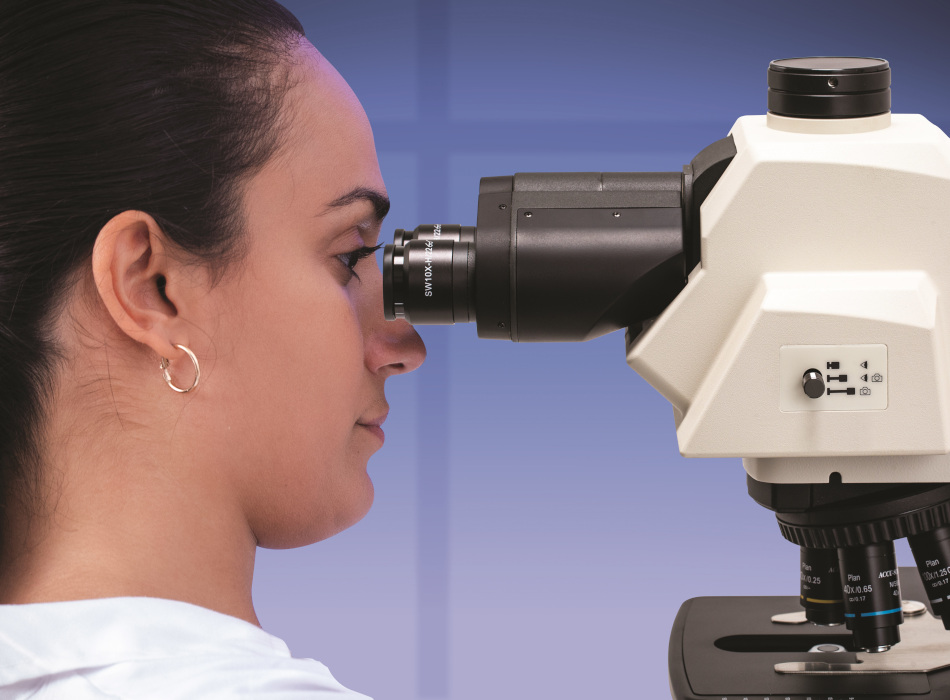
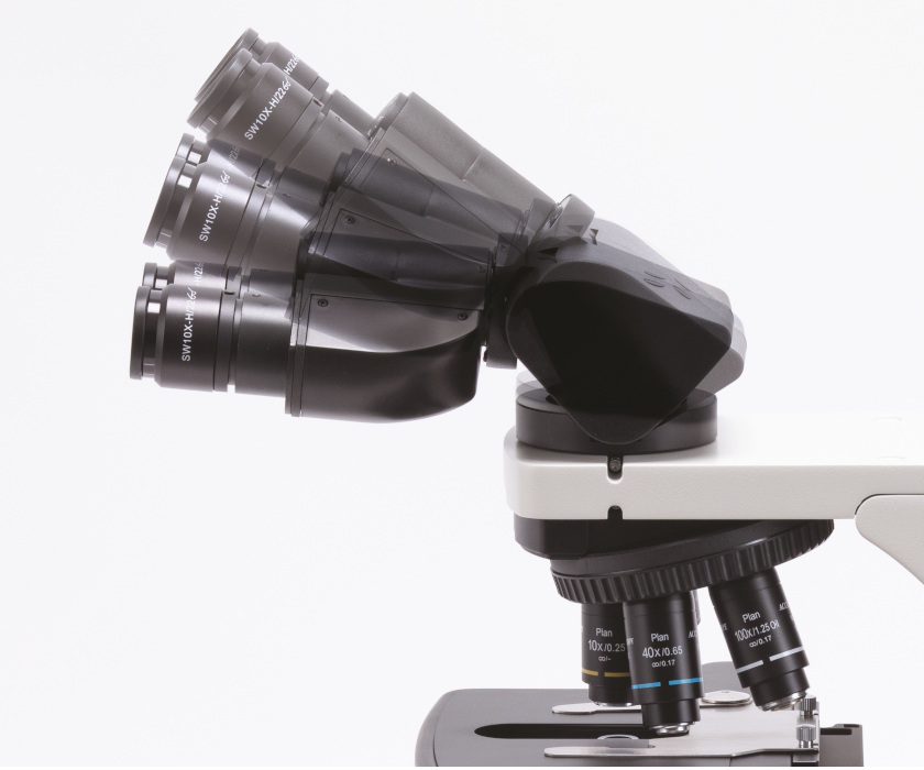
With a choice of an ergonomic binocular or trinocular viewing head, as well as eye-level risers that can raise the eyepiece tubes in 25mm increments the EXC-500 can maximize comfort and performance in the lab.
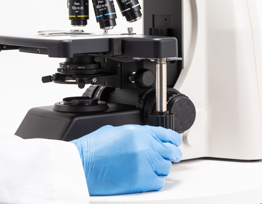
For greater ergonomics, you can combine multiple ergonomic accessories for enhanced comfort. Add a nosepiece spacer to lower your nosepiece and stage by 20mm or 40mm, reducing the strain on your neck, shoulders, and even back during frequent slide changes.
The EXC-500 supports a wide variety of options to visualize your sample through contrast enhancement techniques.
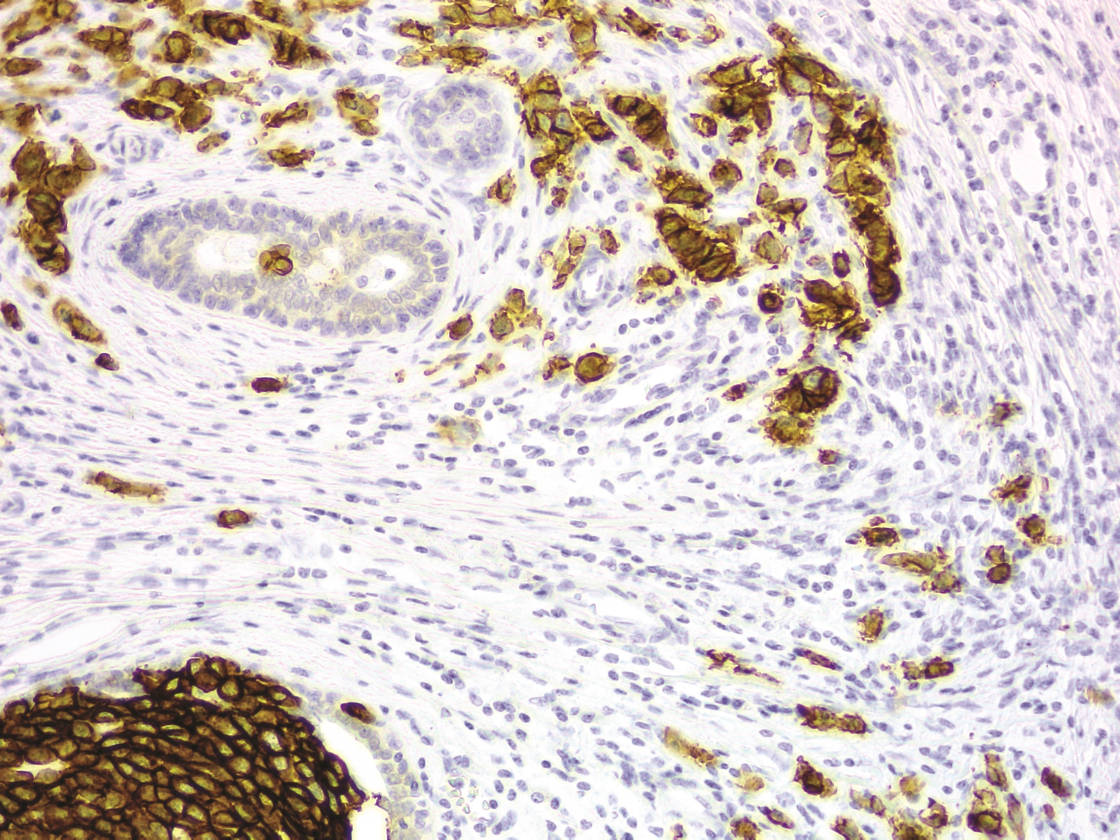
Brightfield:
The most common contrast method in use with upright microscopes, light passes through a specimen with inherent color or that has been stained to highlight structures of interest. All EXC-500 microscopes come standard for brightfield microscopy.
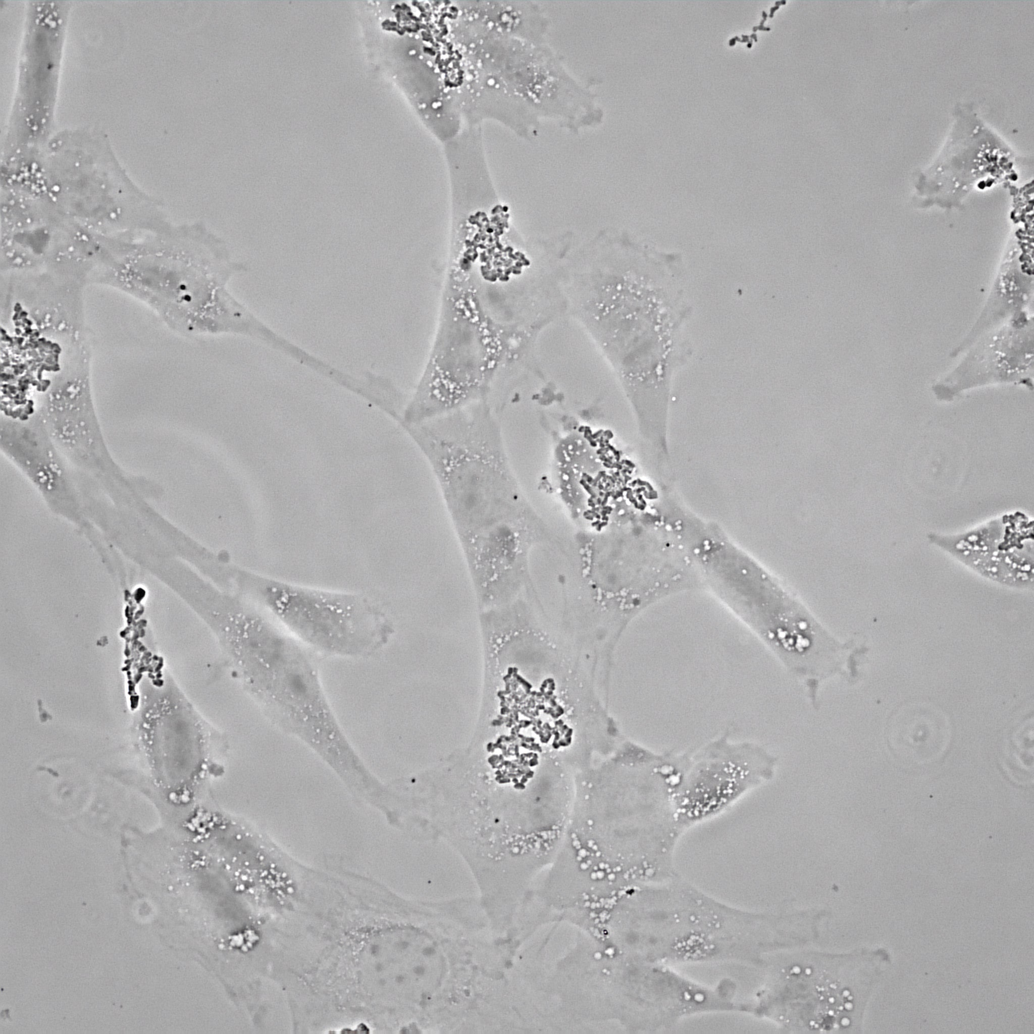
Phase Contrast:
View high-contrast images in unstained samples. Provides additional detail or structural highlights in stained samples.
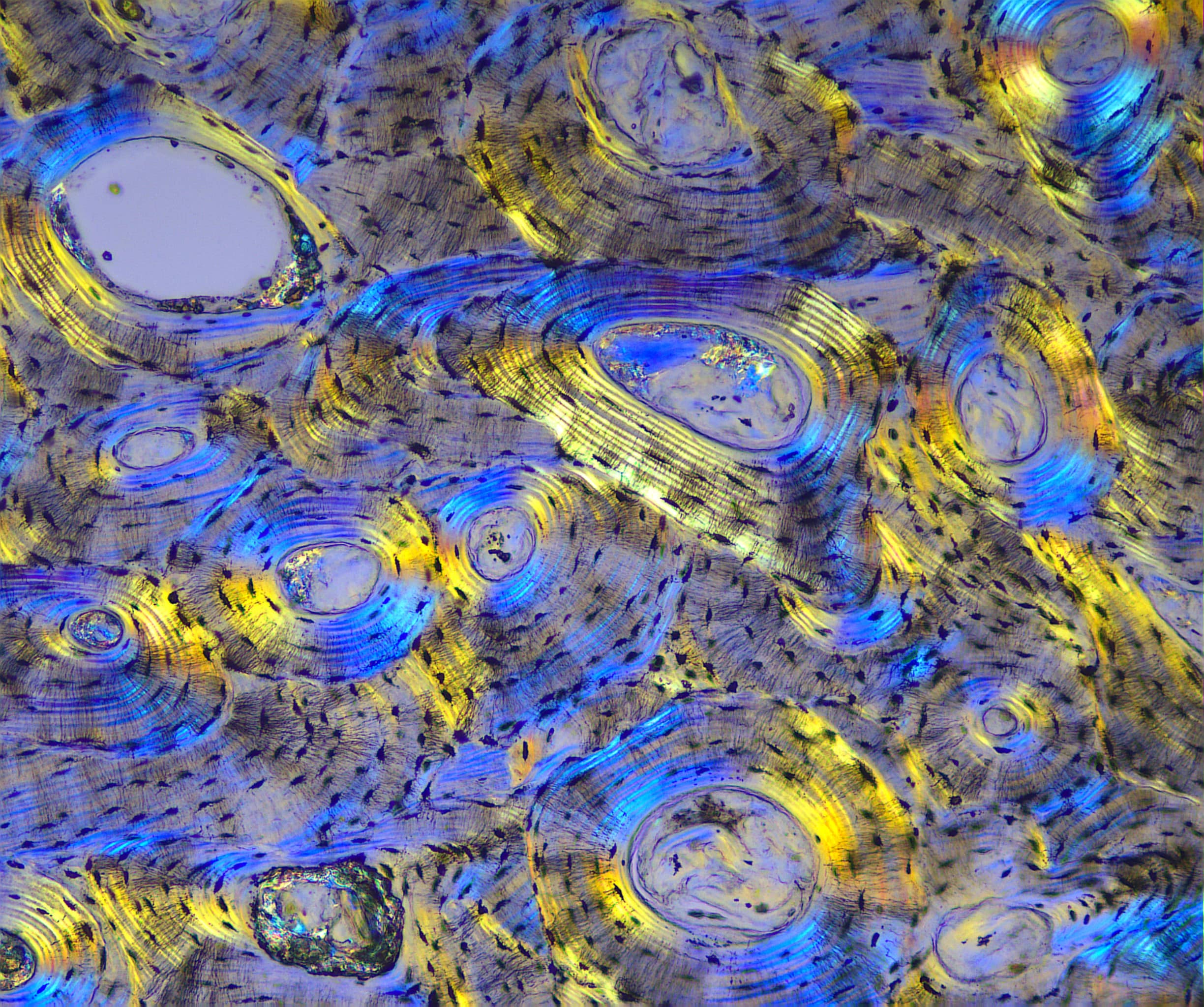
Simple Polarization:
Reveals detail and structures based on optical properties of molecules in the sample. Polarized light microscopy can be used with stained and unstained samples. Gout analysis is a specialized polarized light technique.
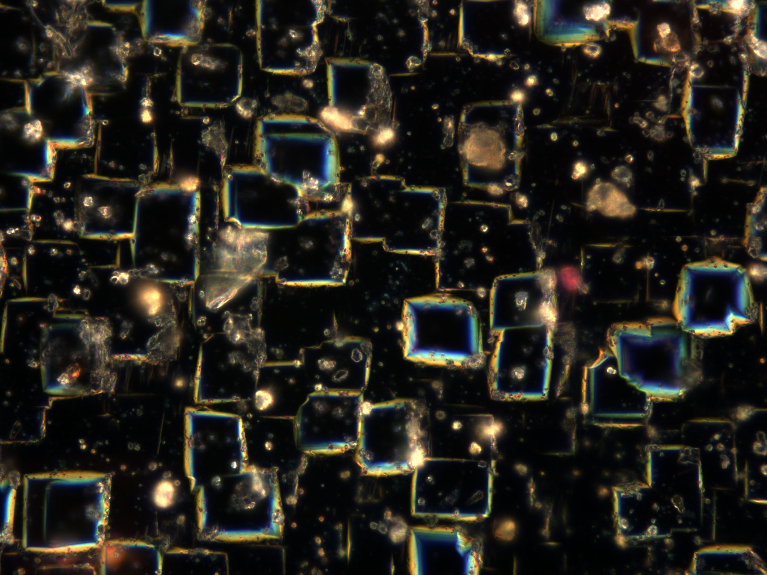
Darkfield:
Utilizes oblique illumination to generate contrast in specimens, resulting in a brighter specimen viewed against a dark background. Use with unstained or stained samples.
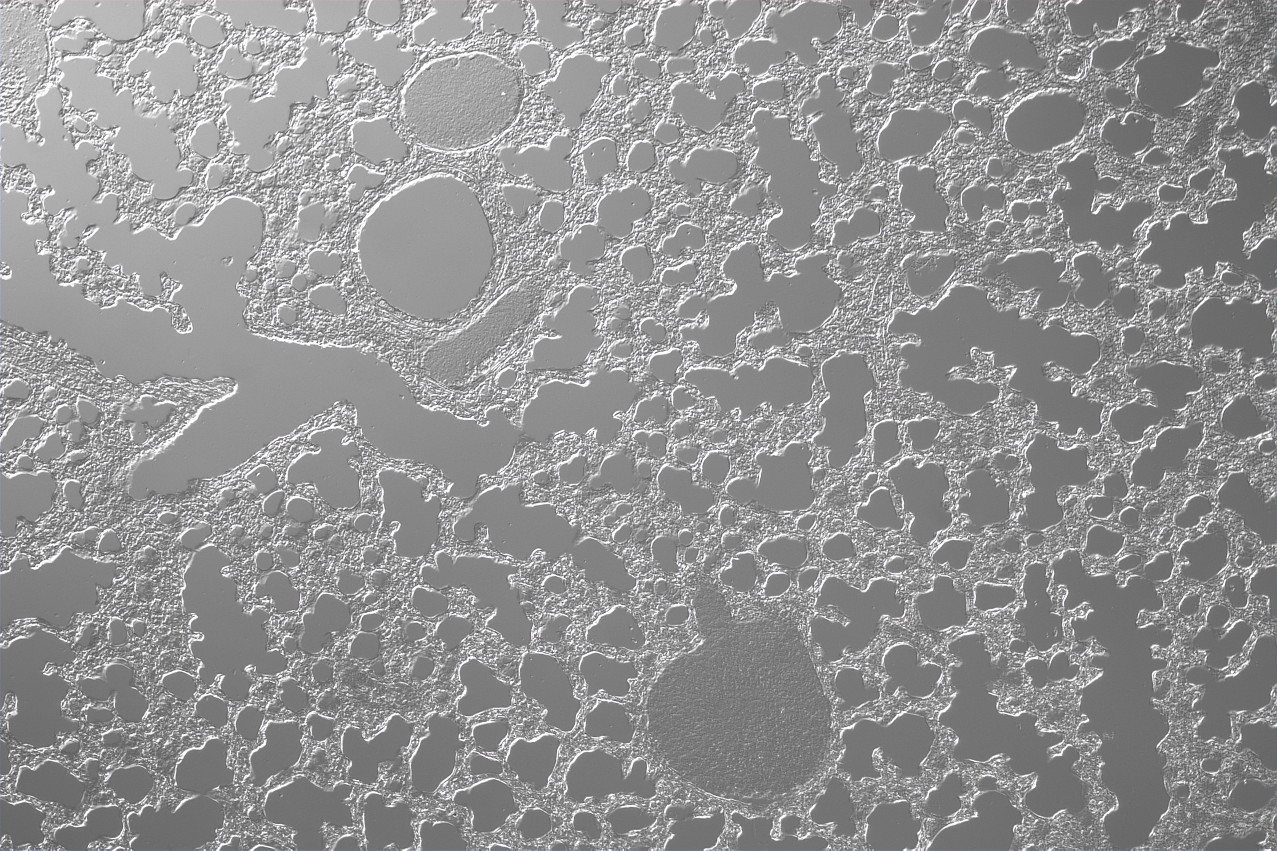
Differential Interference Contrast (DIC):
Creates contrast using differences in optical properties (birefringence) of structures and molecules in the specimen. DIC is used with live and fixed samples, unstained or stained.
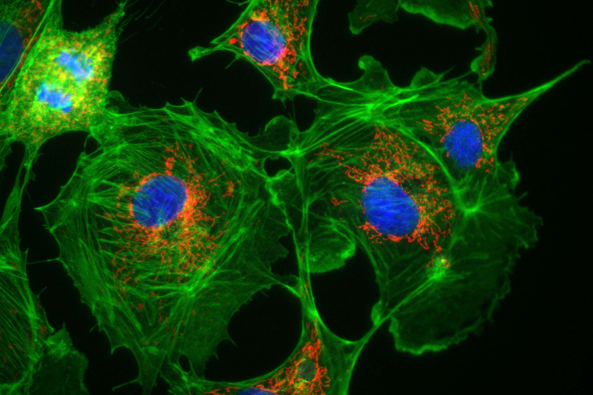
Fluorescence:
View details in specific structures using fluorescent molecules. Structures are targeted using fluorescent dyes, stains, antibodies labeled with fluorescent molecules, fluorescent proteins, and more.
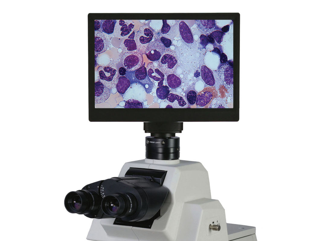
Add a camera for documentation, consultation, or training. Our Excelis 4K and Excelis HD Cameras allow for connection to an integrated 4K or HD monitor or display simultaneous output to an external monitor and PC. Teledyne Lumenera INFINITY series cameras provide excellent performance and offer options such as USB connectivity and HDMI output. Our SKYE WiFi cameras offer increased flexibility with a combination of USB, Ethernet, and HDMI for enhanced connectivity. All our cameras come with FREE software!
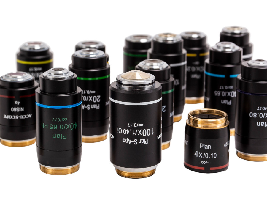
Quality images begin with the optics. ACCU-SCOPE’s NIS Optical System offers a wide range of Plan Achromat, Apo, Apochromat, and Plan Phase objectives to meet the performance requirements of many contrast methods.
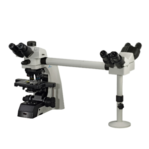
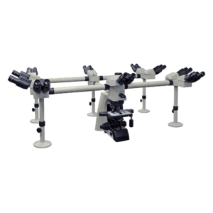
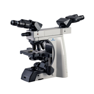
The EXC-500 can support multiple observers from 2 to 10. Add a camera to extend sharing and instruction capabilities beyond the microscope.


Choose from a variety of accessories to adapt your EXC-500 for your work today, and your work in the future.
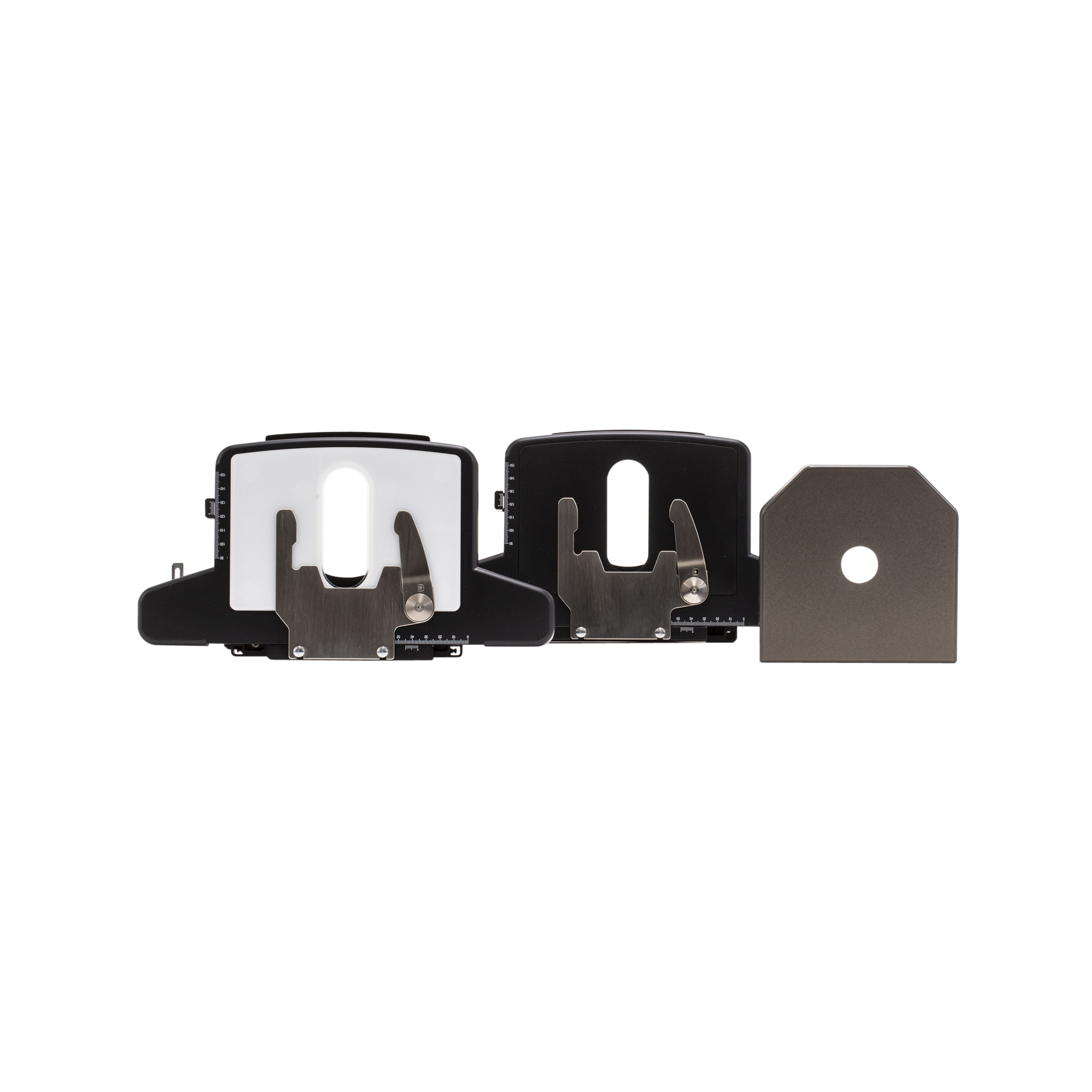
Choose the stage that fits best with your workflow. Our hard-coated white Gorilla™ glass stages are available in right- or left-handed versions. Our special pathology stage features a ceramic coating and no slide holder for users who prefer to manually position the slide on the stage.
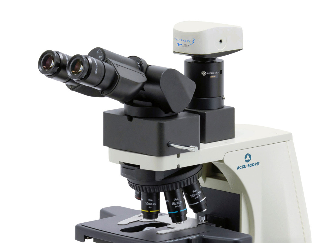
Upgrade your EXC-500 with a camera, even if it has a binocular viewing head. Simply supplement with our trinocular viewing accessory (requires camera and camera adapter) and keep using your current viewing head. You can also change to a trinocular head, then add a camera and adapter.
"*" indicates required fields