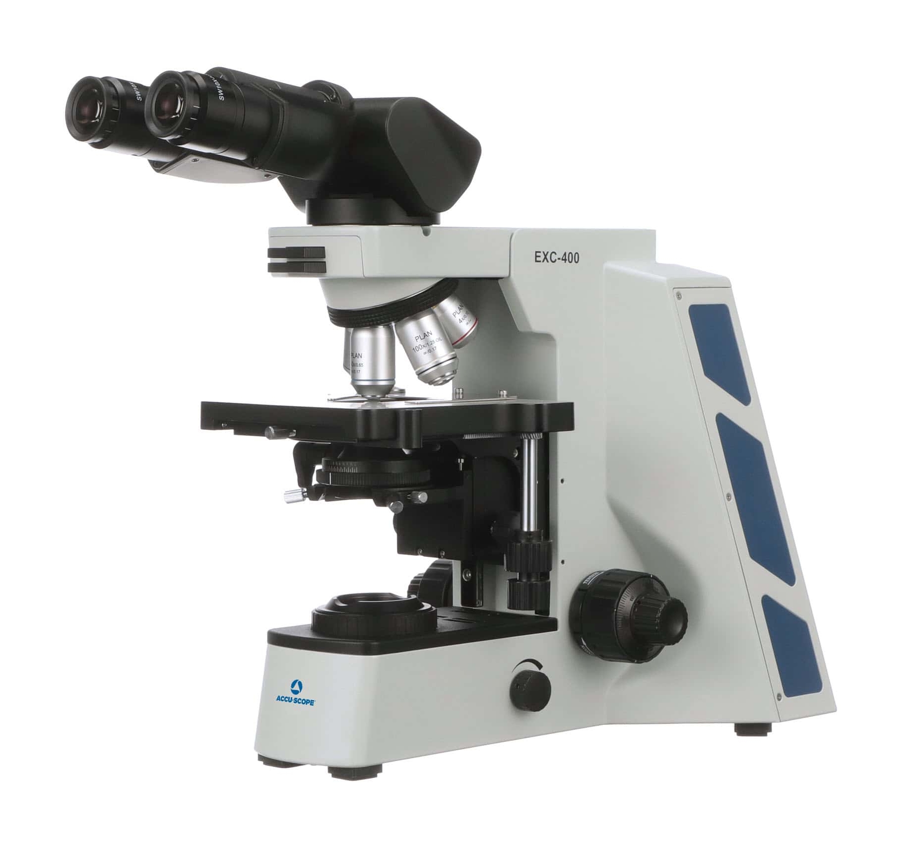
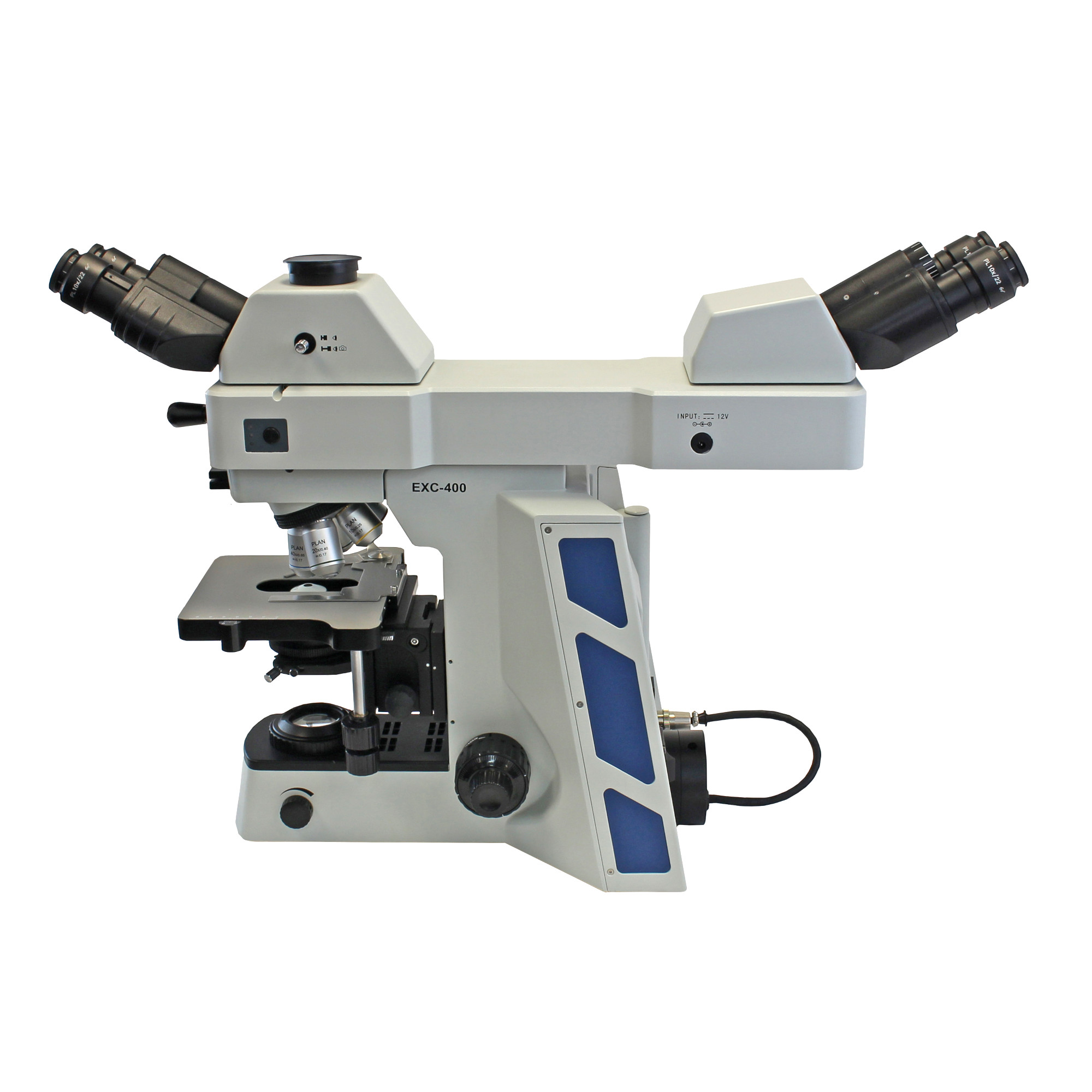
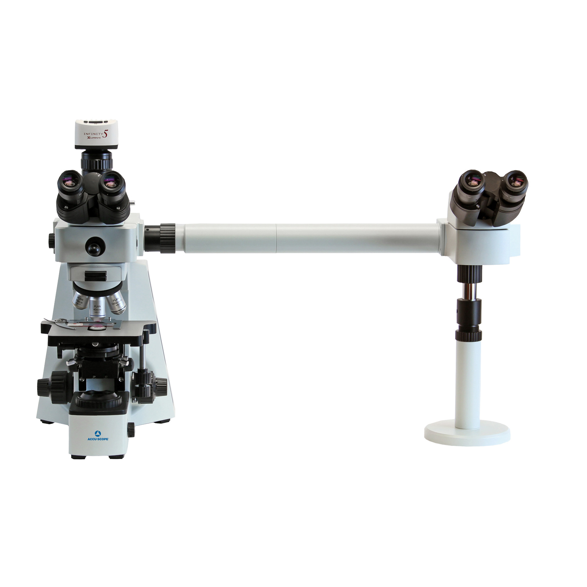

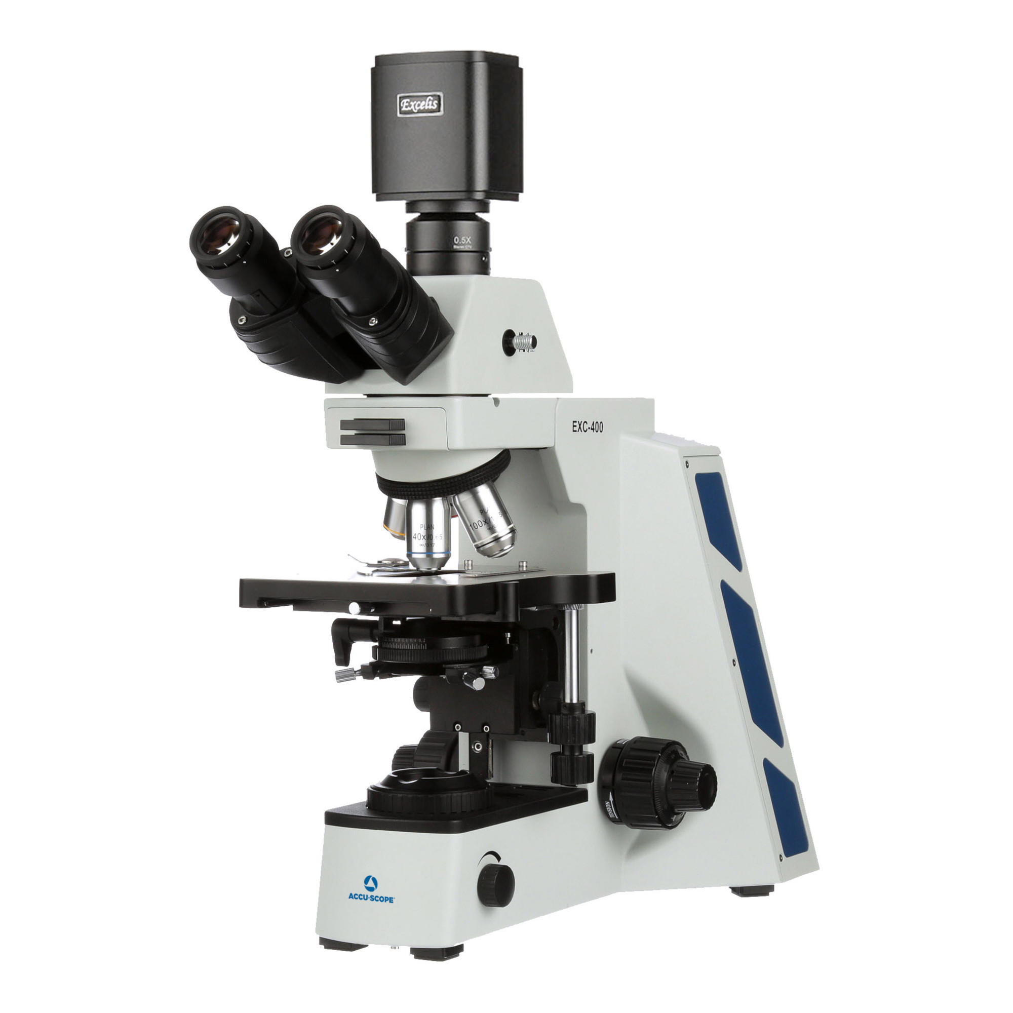
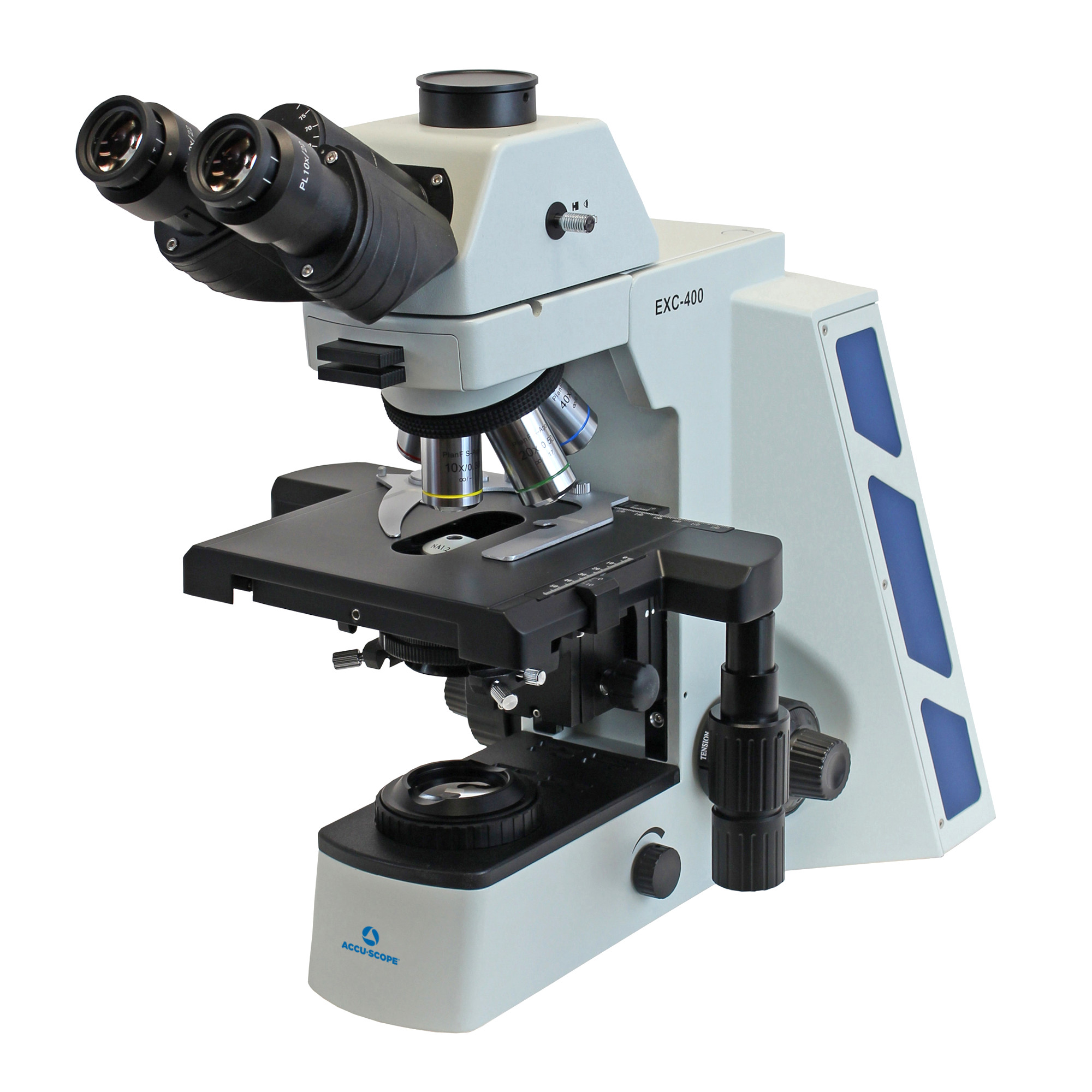
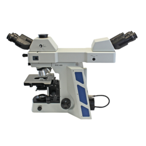
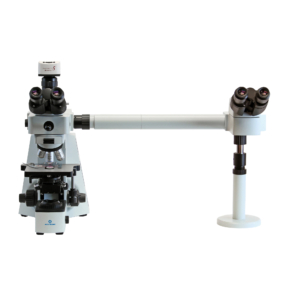
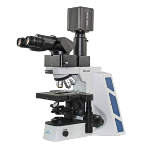
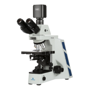
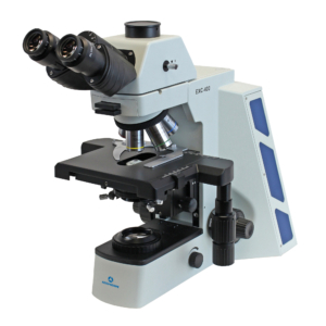
The EXC-400 from ACCU-SCOPE is designed for a broad range of microscopy applications in clinical, academic, and research environments. The EXC-400 is ideal for routine observations and can be configured with several contrast methods to meet your application needs including brightfield (standard), phase contrast, polarized light, gout analysis, and fluorescence. Choose a trinocular or ergonomic binocular viewing head, or one of our dual-viewing options (available as side-by-side and front-to-back configurations). The broad triangular footprint makes the EXC-400 exceptionally stable on any bench surface.
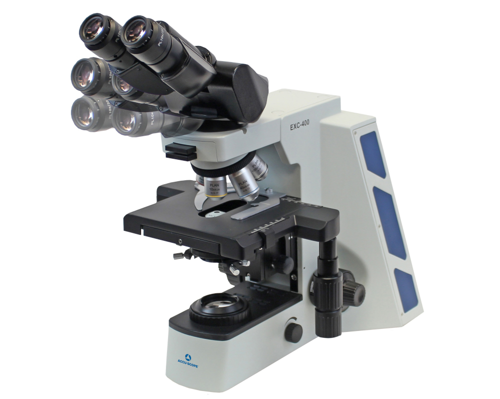
Using the ergonomic binocular viewing head, users can incline the eyepieces between 5˚ – 35˚ to allow for natural viewing posture during sustained use periods. An optional camera port can be added to allow for digital imaging and documentation. The eco-friendly LED illuminator provides consistent, color temperature throughout the magnification range offering uniform brightness and user comfort.
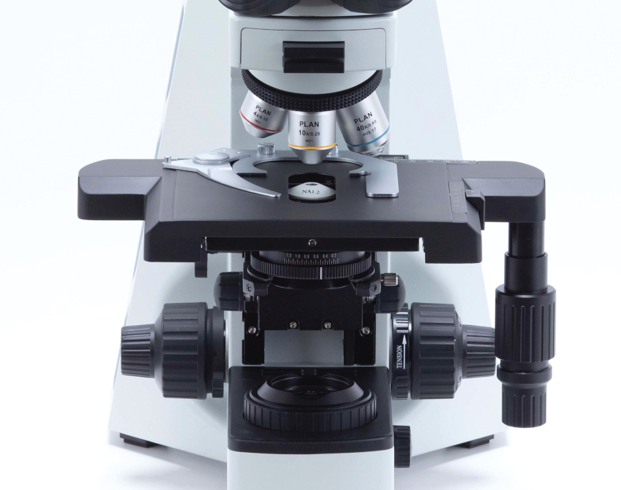
The scratch-resistant, coated stage with adjustable tension controls allows the user to set the tension of the stage controls allowing users to easily customize the movement feel of the stage controls to maximize individual comfort. The coaxial controls are conveniently located near the focus knobs and low towards the bench surface allowing the user to rest their hand on the counter while adjusting the stage in either direction.
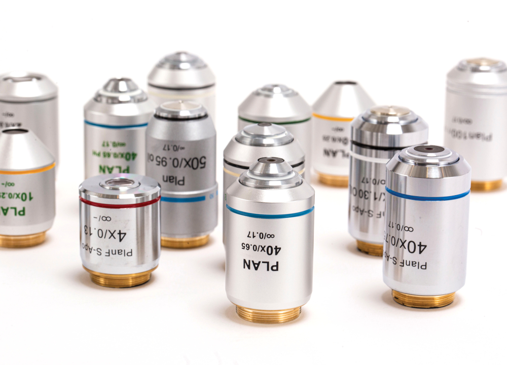
Defined by superb clarity, sharpness, and resolution, the EXC-400 objectives provide bright images for routine and challenging laboratory observation work. With a wide range of objective lenses ranging from Plan Achromat to Semi-Apo to Plan Phase lenses, the EXC-400 Series is a modular choice for today’s discerning laboratory.
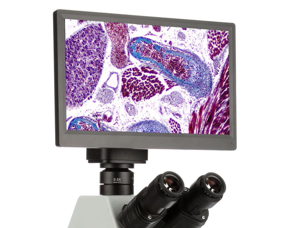
Offering a wide range of low-profile camera adapters, the EXC-400 Series can accommodate a wide range of camera choices for all applications. ACCU-SCOPE’s Excelis cameras offer best in class 4K, HD and USB camera options with the ability to integrate space saving 4K and HD monitors onto the camera.
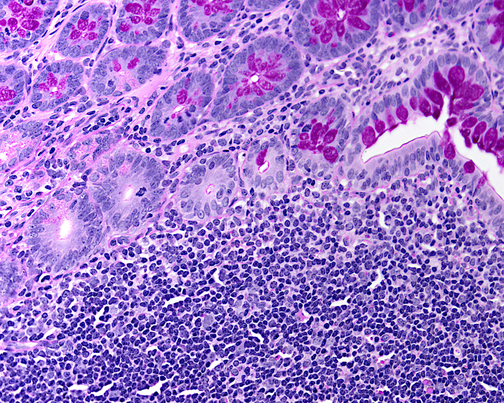
Brightfield – The most used contrast method in microscopy is brightfield. Light passes through a specimen that has been stained or has inherent coloration that highlights features of interest. All EXC-400 microscopes come standard for brightfield microscopy.
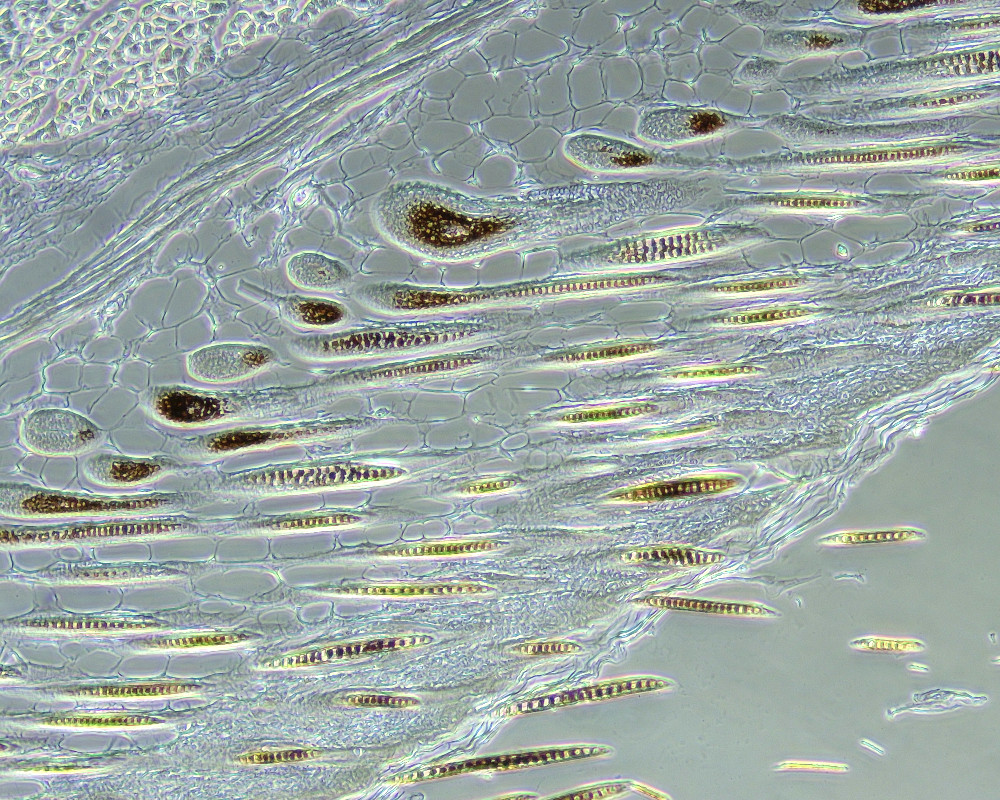
Phase Contrast images can be observed with the optional turret phase condenser and plan phase objectives. Phase contrast can be used with unstained or stained samples.
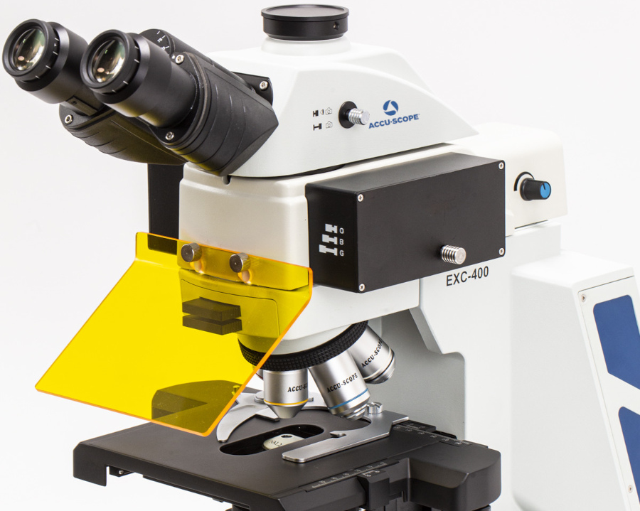
With either a 6-position fluorescence filter wheel that can accommodate up to 6 filter cubes or integrated single, dual or three channel LED fluorescence illuminators, the EXC-400 can provide high performance fluorescence imaging for even the most challenging specimens.
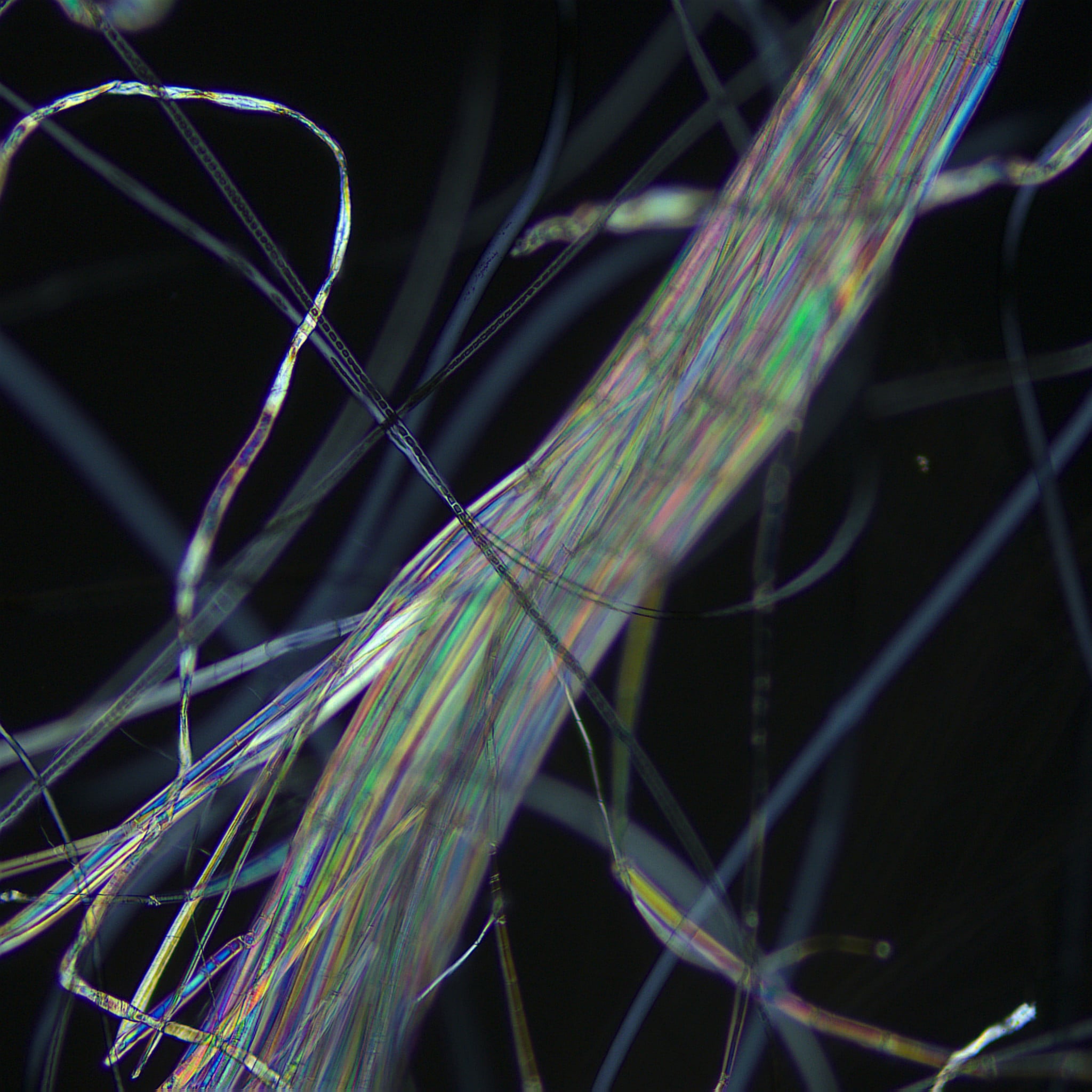
Ideal for viewing birefringent substances such as crystals, cellulose in plant cell walls, starch grains and certain tissues (e.g., collagen in fibrosis).
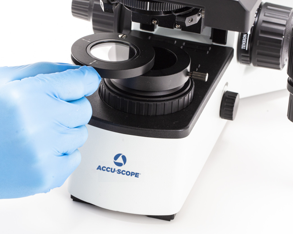
The optional gout analyzer allows for the identification of uric acid crystals by changes in the interference color.
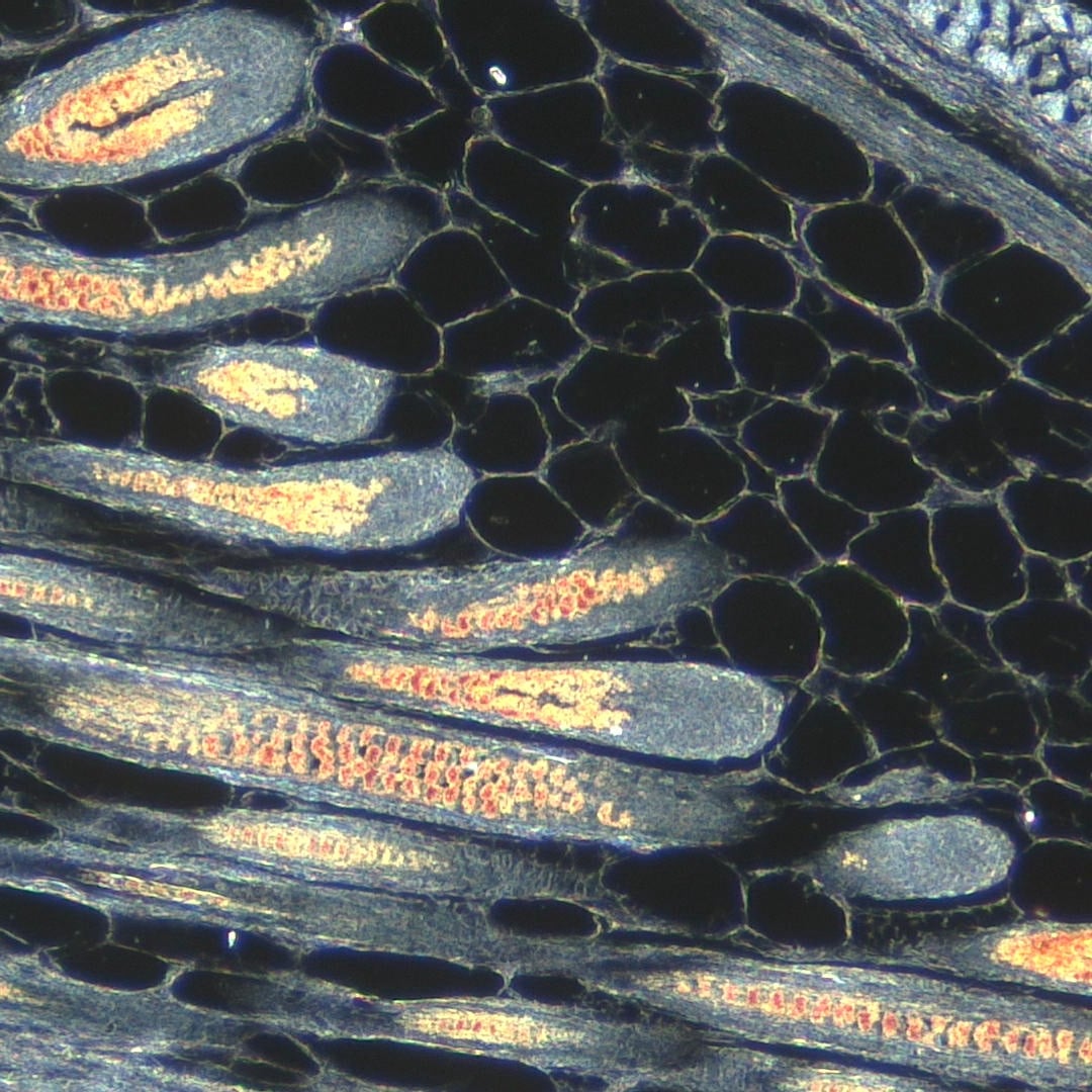
Available as an option on our turret phase condenser, a darkfield filter (annulus) delivers light at an extreme angle to the specimen. In the absence of a sample, the light passes by the objective and the field of view is black. Only light striking components in the sample will be scattered, some of which is captured by the objective and seen as bright objects against the otherwise black background. Darkfield allows for the observation of unstained samples, living or fixed.
"*" indicates required fields