Phase contrast microscopes utilize a special technique to enhance the contrast of an image. This technique was developed to produce high-contrast images of transparent or unstained specimens that could not be viewed well under brightfield illumination. Here, we take a look at some of the advantages offered by phase contrast microscopes for clinical settings and other fields that frequently work with living cells, microorganisms, and similar specimens.
Observe Living Cells
Before Dutch physicist Frits Zernike invented the phase contrast microscope in the 1930s, scientists were unable to clearly view living cells. Instead, the organisms were dried on a slide and stained in order produce contrast. However, this process takes time, kills the cell or organism, and can lead to a loss of structure and, therefore, detail. By being able to view an organism while still living, scientists can see how the organism behaves, offering further insight.
View Thin or Transparent Specimens
Specimens that are transparent or translucent, such as very thin slices of tissue, can be difficult to study using brightfield, as much of the detail remains hidden to observation. Phase contrast can make it easier to view and study the details of the internal structure of a cell or microorganism thanks to the increased contrast and minimal manipulation (e.g. fixing) of the specimen.
Can Be Combined with Other Observation Methods
To get the best possible image for their specimen or application, scientists sometimes need to experiment with different observation methods. Phase contrast can be easily combined with other methods of observation, such as fluorescence, to reveal additional detail that couldn’t be observed with just one method.
Applications of Phase Contrast Microscopes
Phase microscopes are not limited to clinical applications. Many different types of specimens can be easily viewed with a phase contrast microscope, including:
- Live microorganisms, such as bacteria, mold, protozoa, and erythrocytes
- Fibers
- Lithographic patterns
- Glass fragments
- Subcellular particles, such as nuclei and other organelles
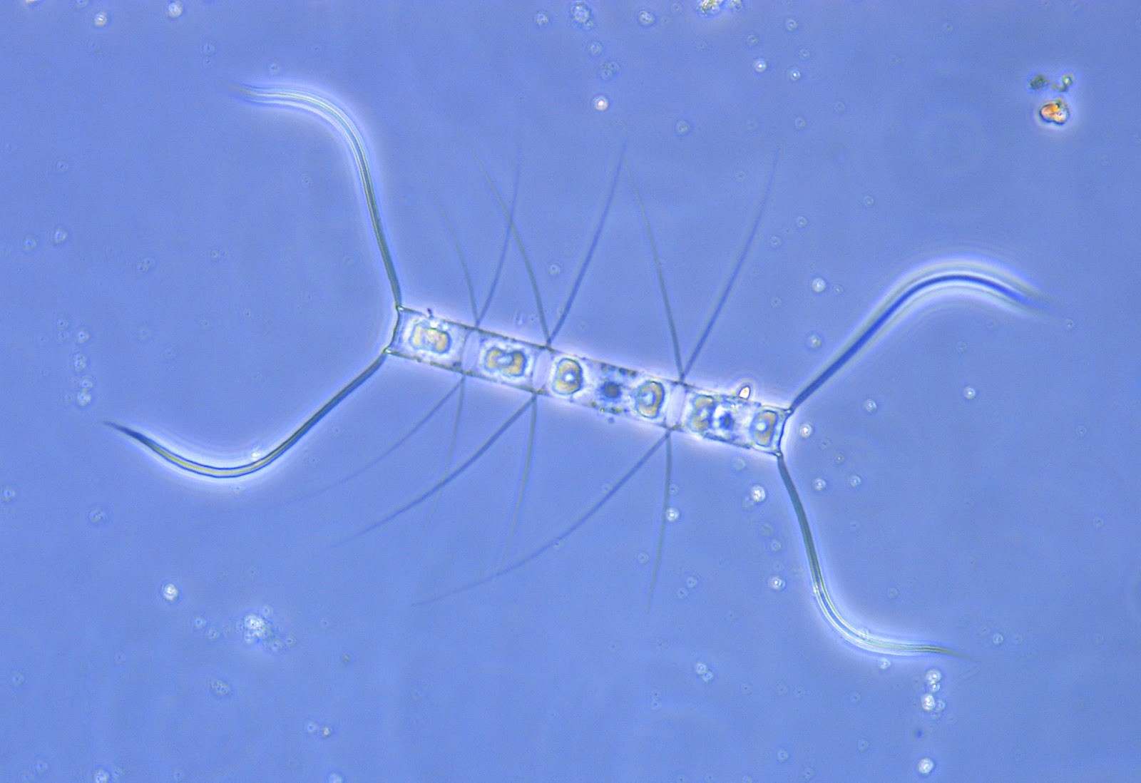
Phase contrast produces a unique “halo” around the edges of particles. This hallmark of phase contrast microscopy is particularly useful to rapidly locate tiny specimens in the sample such as bacteria or debris.
Find Phase Contrast and Other Specialized Microscopes from ACCU-SCOPE
No matter your laboratory’s requirements, we can help you choose the right microscope that fits your needs and budget. ACCU-SCOPE offers microscopes for clinical settings and many other specialized applications. To learn more about our products, or for any other inquiries, please reach out to us online or by phone.
