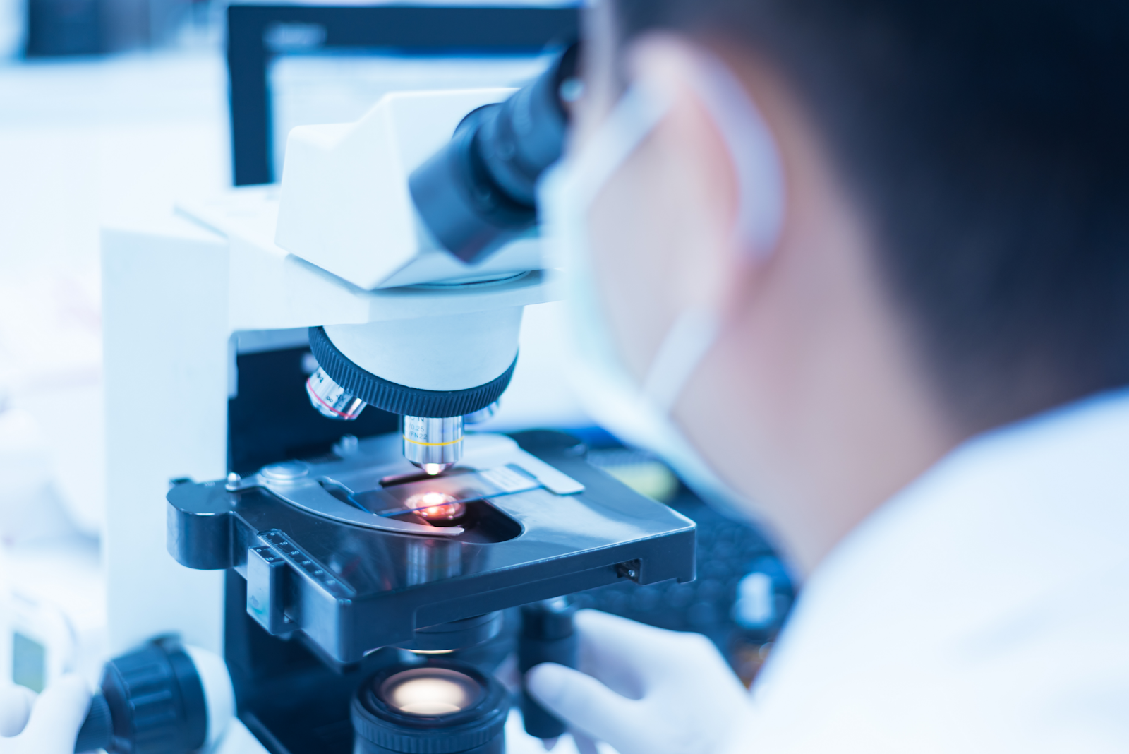
The term microscope originates from the Greek words mikros meaning “small,” and skopein, meaning “to look.” The first recorded use of a microscope-like instrument dates back to the late 16th century. Yet, it wasn’t until the mid-17th century that significant advancement in magnification enabled Van Leeuwenhoek to observe what he called “animalcules” in samples. These “animalcules” were later renamed bacteria and found to be the cause of several diseases such as tuberculosis and the plague. Still, years would pass before viral agents could be successfully observed with microscopy due to past limitations in magnification.
During the latter part of the 19th century, Adolf Mayer speculated that an unknown and unseen infectious agent was causing mosaic disease in tobacco plants. Although he never physically viewed the virus, further experiments involving the filtration of plant particulate matter showed the presence of an undetermined pathogen. It would take decades for the tobacco mosaic virus to be identified in crystallized specimens leading to the additional discovery of over 900 viral variants that infect plant species.

Early Discoveries in Microscopy
In the mid-20th century, the development of the electron microscope enabled researchers to finally observe viruses. By utilizing accelerated electrons, scientists were able to see particles significantly smaller than any elements viewed through the optical microscopes of the time. Although the visual discovery of viral particles was a profound advancement in observational methods, the inability to see viruses work in real-time using an optical microscope would remain elusive until the end of the century.
During the 1980s, light-based observation of viruses was finally possible using highly sensitive microscopes. For the first time, researchers could see viral particles interact with one another and inside cellular membranes. The mechanics of an infection caused by a virus could now be viewed in real-time, enabling scientists to better understand mechanisms of viral infection and illness. The virus could no longer elude detection or the new treatments that would be discovered as a result.

Modern-Day Microscopy
Significant leaps in technological advancements continue in the field of microscopy today. With advances such as phase-contrast microscopy, which can be used to study the impact of viruses on living cells, to TIRF microscopy, which can increase the effectiveness of detection utilizing specialized dyes, the field of microscopy is continuing to open the doors of scientific discovery.
ACCU-SCOPE is a leading provider of microscopes and accessories to provide researchers with robust detection capabilities. The pursuit of knowledge through the scientific process combined with technological advancement paves the way for a brighter future. Through microscopy, we will all benefit from a better understanding of viruses and their impact on us and the environment. To learn more about our microscopes for clinical and research laboratories, please feel free to contact us today.
