

It is believed that compound microscopes (also known as upright microscopes) have been around since 1590, when Dutch spectacle-maker Zacharias Janssen invented this incredible piece of technology. Since then, scientists and students alike have used upright microscopes to examine specimens that are too small to be visible with the naked eye. What sets them apart from other microscopes is their use of multiple lenses for magnification, flatness correction and color correction. Typical upright microscopes have objective lenses and an eyepiece lens to obtain magnifications between 40x and 1000x. These microscopes are often the go-to choice thanks to their incredible power and simplicity in operation.
To determine if an upright microscope is right for you, let’s look at how it works and in which applications it is best used.
Parts of an Upright Microscope
The components of your particular upright microscope may vary based on the configuration of the instrument, but most uprights have the following features:
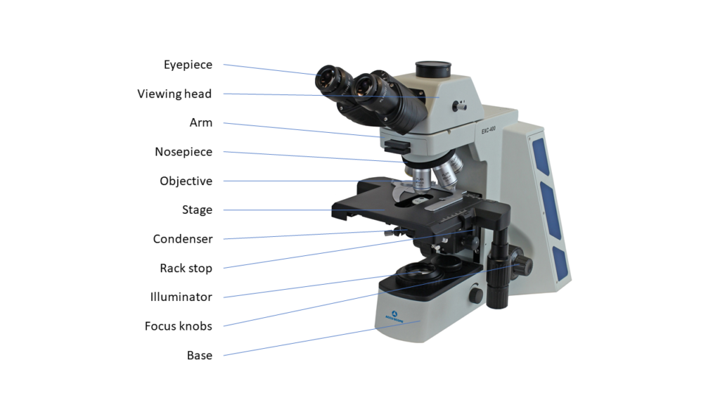
- Eyepiece (ocular): This is the lens through which the observer will look. It typically provides 10x or 15x power.
- Viewing head: This is the connector piece between the eyepiece and the objective lenses.
- Arm: This piece connects the head to the base of the microscope.
- Base: This is the bottom of the microscope.
- Revolving nosepiece: This part of the microscope holds the objective lenses. Rotate the nosepiece to change to another objective..
- Objective lens: Typically, an upright microscope will have three or four objective lenses. The shortest lens has the lowest power, while the longest objective usually has the highest power.
- Stage: This is the platform where the observer places their slides. The stage usually has clips to hold the slides in place. If the stage is mechanical, two knobs on the side will allow the stage to move left and right.
- Condenser: This component is located under the stage (between the sample and illuminator) and concentrates the light delivery to the specimen.
- Rack stop: This component determines how close the objective lens can get to the slide. It is typically set in the factory to prevent the lens from coming down on the slide and breaking it.
- Focus knobs: This component moves the specimen to be in focus through the eyepieces by moving the stage up or down. A coarse focus knob moves the stage faster, while a fine focus knob allows for precise focus of the sample.
- Illuminator: This is the steady light source shining up through the stage.
How an Upright Microscope Works
Unlike a stereo microscope where the light source usually shines from above onto the specimen, an upright microscope’s light source is transmitted from below the stage and upwards through the sample. The observer then looks down upon the specimen through an ocular lens. The focus knobs adjust the object up or down to be in focus to the eyes. The power of the eyepiece and objective lenses are multiplied together thereby creating greater magnification than a single lens alone. Because the working distance between the objective and the sample is so small, upright microscopes are ideal for thin or flat subjects like bacteria on a slide rather than something that is thicker (e.g., a housefly) or that needs to be dissected.
When to Use an Upright Microscope
Upright microscopes are used to observe small samples or details that are generally too small to be seen with the naked eye. They are ideal for life science and cell biology applications because they can utilize basic observation methods including brightfield, phase contrast, darkfield, and fluorescence microscopy of samples (see examples below).

To reveal greater detail, an upright microscope is often used to view thin slices of larger tissue samples from animals. Brightfield observation uses the inherent color in a sample or samples that are stained to add color to and enhance certain features. Phase contrast uses an optical technique to enhance contrast to usually unstained samples, and some existing color may be lost in this process. In darkfield microscopy, light that strikes a sample is scattered and some is captured by the objective for observation, whereas unscattered light never gets captured, resulting in a dark background. Fluorescence microscopy uses fluorescent molecules to tag particular structures in a sample, making them visible to the eye. Differing from the other observation methods above, the light source for fluorescence microscopy shines from above the sample. Because of their ease of use and powerful magnification, upright microscopes can be readily found in various clinical, educational, pharmaceutical, research, and veterinary laboratories.
Trust ACCU-SCOPE to Meet Your Upright Microscopy Needs
Since its invention over 400 years ago, the upright microscope has been used to make amazing scientific discoveries, and is still the most recognized and popular choice of microscope for viewing specimens on a slide. If you’re shopping for quality microscopes for your lab, turn to ACCU-SCOPE for all your microscopy needs. We have an array of microscopes and accessories, including upright microscopes and microscope heads. If you need low magnification for basic observation and dissection, we also have information on stereo microscopes. Don’t hesitate to contact our team to determine which type of microscope is best for your application.