For centuries, biologists have been curious about the development of embryos. Because of the advancements in microscopy, our understanding of embryogenesis has not only evolved but now some of our best medical professionals use optical technologies to help women become pregnant.
When a woman and her partner struggle to conceive, they often turn to assisted reproductive technologies (ART) to achieve pregnancy. ART treatments include in vitro fertilization (IVF), intracytoplasmic sperm injection (ICSI), and intracytoplasmic morphologically-selected sperm injection (IMSI). Each method aims to fertilize a female’s egg outside of her body. For the procedure to go smoothly, the IVF laboratory must have high-quality microscopes to support ART.
Ways Microscopes Can Aid Assisted Reproduction
There are several reproductive medicine approaches that use different microscopy techniques to enhance a woman’s ability to become pregnant. These reproduction techniques are IVF, ICSI, and IMSI. Before fertilization can occur, an IVF scientist must take steps to prepare the sperm and egg. Each of these steps requires the use of microscopes. Let’s take a closer look at these procedures:
Semen Analysis
Before artificial fertilization can occur, an experienced scientist evaluates the quality of the sperm. They typically use an upright microscope to determine the total number, motility, and morphology of the sperm. Only healthy sperm will be used for fertilization. This pre-selection process requires a microscope with DIC or polarized light illumination.
Oocyte Preparation
After extracting oocytes from the female patient, the outer egg cell layers (besides the zona pellucida) are removed. This process is called “denuding” and is performed in a Petri dish while observing under a stereo microscope. The egg is transparent, so excellent illumination and contrast control are essential to be able to see the egg and its layers. Diascopic stands are the stereo microscope stands of choice and feature a tilting mirror to angle the light to provide oblique or darkfield contrast. Once this step is complete, the IVF scientist evaluates the morphology of the oocyte using an inverted microscope for its high magnification and easy accessibility to the egg for micromanipulation. If abnormalities are detected, the oocyte will not be used during fertilization.
Fertilization
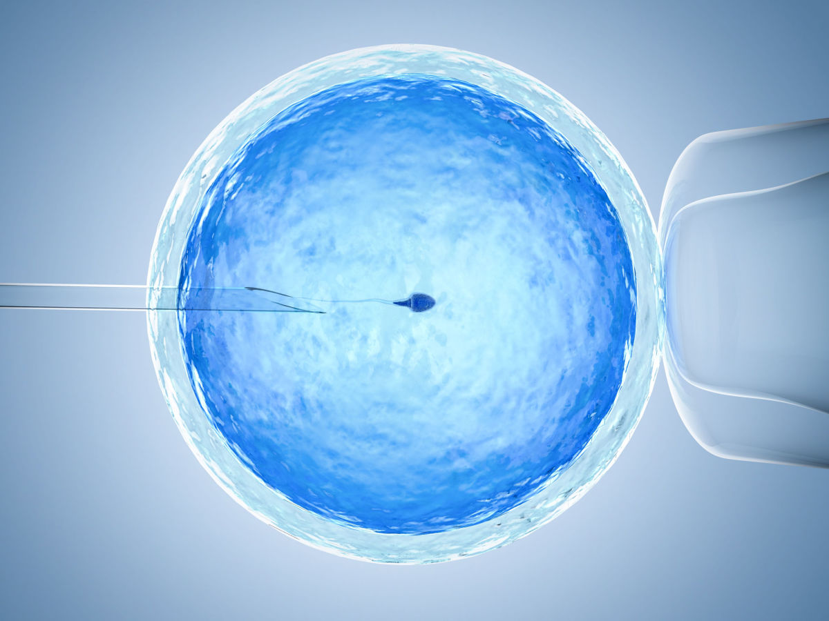
Once the healthy sperms and oocytes have been isolated, artificial fertilization follows next. With ICSI, a single sperm cell is injected into an oocyte using a micromanipulator. The oocyte’s zona pellucida and polar body must be visible for this procedure. An alternative to ICSI is IMSI, which involves the extra steps of assessing sperm morphology using an inverted microscope and injecting sperm with the desired morphology.
Embryo Growth
After the eggs are fertilized, an embryologist monitors the growth and development of the embryos over the next few days. They look for any imperfections in the embryos. Only the best and healthiest embryos are implanted into the mother.
How to Choose a Microscope for IVF Applications
ART laboratories require a range of microscopes to complete various clinical procedures, such as gamete selection and embryo monitoring. Choosing the right microscope requires considering several factors, including the specific operation or application and the microscope’s features. When selecting a microscope for IVF applications, look for the following features:
Stereo Microscope
- A zoom range that provides enough magnification to see detail in the egg
- Diascopic stand that generates the necessary contrast to reveal the detail in the egg
Inverted Microscope
- An inverted design that allows access to the cells or embryos for manipulation
- A range of objectives to ensure sufficient magnification that facilitates observation of the specimen and the use of micromanipulation tools during sperm and egg cell handling
- A built-in camera port to support the use of a digital camera for viewing specimens and documentation
Need Microscopes for IVF Procedures? Turn to ACCU-SCOPE
IVF laboratories need high-quality microscopes they can rely on. If you’re searching for stereo, inverted, and upright microscopes to outfit your IVF and embryology laboratory, you’ve come to the right place.
At ACCU-SCOPE, we have a wide selection of microscopes used for embryology and IVF, including stereo and inverted microscopes with cameras. We can even equip your lab with stereo stands and accessories that work with various microscope models.
Contact our team today for help choosing the right microscope for your application!
