By now you understand if you need a color or monochrome camera, and you've determined how you will attach the camera to your microscope. But what about the “megapixels”? More is better, right? If you are using a microscope as your camera lens, our recommendation is not to follow the general consumer hype that more megapixels are better. Although this may be true for your cell phone, the logic doesn’t transfer to your microscope. Let’s look at resolution more closely from the perspective of microscopy imaging. Warning: This may get a little geeky.
Unfortunately, choosing the optimal camera resolution isn’t as easy as picking color or monochrome. This will involve a little bit of math and in the end, you will have a general idea of when more resolution is better and when it’s not.
Optimal camera resolution (pixel size and # of pixels) depends on the resolution of the microscope and the magnification that is used for imaging. In a microscope two objects are said to be “resolved” when they can be clearly distinguished from each other. This is referred to as the Rayleigh Criterion – it defines the optical resolution of the microscope and is illustrated below. When viewing objects in a microscope that are below the limit of resolution (we refer to them as point sources of light), each creates a pattern called an Airy disk, appearing somewhat like concentric ripples in water with a much taller peak in the middle. Figure 6 below illustrates a sideview of the Airy disk pattern of two objects that are fully resolved, just resolved, and not resolved, respectively.

Mathematically, the Rayleigh Criterion (d) is described as:
d = 0.61λ/NA
... where λ is the wavelength of light and NA is the Numerical Aperture of the objective (this assumes that your microscope has been correctly adjusted for Köhler illumination, so the condenser has the same NA as the objective). Why does the wavelength of light matter? Simply stated, resolution improves with shorter wavelengths -- better resolution with blue light than red.
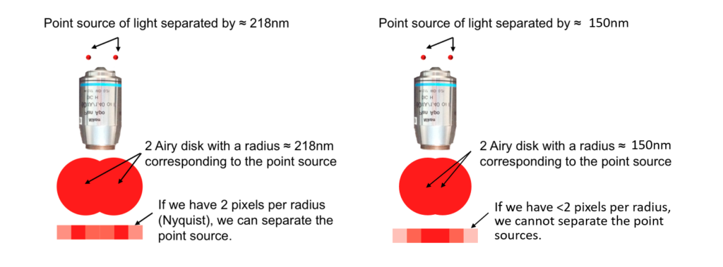
In microscopy, Rayleigh’s criterion is typically expressed in nm (nanometers) or µm (microns, micrometers). So how does this fit with the number of pixels in a camera? The Nyquist-Shannon theorem requires a sampling interval (in our case the pixels of the camera) that is at least twice the optical resolution. You can also think of the Airy disk of each object being covered by at least 2 pixels. Consider that each of the two objects mentioned above are so small that they are only detected by one pixel each – their size is at or below the optical resolution of the microscope. If the objects are detected by adjacent pixels, we couldn’t tell if it were two objects or one slightly larger object. But if there is an empty pixel between them thereby satisfying the Nyquist-Shannon criterion, then we can say that the two objects are resolved on the camera.
Below is a figure adapted from the Cell Sciences Imaging Facility at Stanford University. It illustrates two objects (point sources) that are separated by the resolution of the microscope according to Rayleigh’s Criterion. The Airy disks for each cover approximately 3 pixels wide on the camera sensor. Therefore you can see that the bright peaks of each Airy disk can be clearly identified by single pixels that are well separated by two other pixels. If we move the objects closer together, you can see that we would not be able to definitely say that our camera can detect two separate objects or just one that may span two pixels.

Magnification adds an additional more variable to the calculation for camera resolution. Higher magnifications project larger images onto the camera sensor, so the pixels can be larger and still satisfy the Nyquist-Shannon criterion. Larger pixels collect more light and, therefore, are more sensitive.
So here is the quick takeaway message about camera resolution. If you are using lower magnifications (e.g., stereo microscopes at lower zoom levels), then you will want smaller pixels and more of them. If you are imaging at higher magnifications (e.g., 40X objective and higher), then you will want larger pixels.
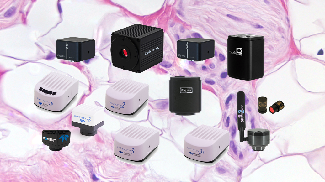
Below are our recommendations for cameras depending on your microscope and magnification. Although they may not exactly fit the criteria discussed above, they are reasonably close and will perform adequately.
Stereo Microscopes:
- BEST:
- Teledyne Lumenera INFINITY8-20. 20MP with USB connection to PC.
- BETTER:
- Teledyne Lumenera INFINITY8-8. 8MP, available in color or monochrome.
- ACCU-SCOPE Excelis 20RC. 20MP with cooling for increased sensitivity and lower noise.
- GOOD:
- ACCU-SCOPE Excelis 20C. 20MP color camera with USB connection to PC.
- ACCU-SCOPE Excelis MPX-6C. 2.4µm pixels.
Upright and Inverted Microscopes (40X objective or higher):
- BEST:
- Teledyne Lumenera INFINITY3 series cameras (4.54µm - 6.45µm pixels, depending on model; color or monochrome)
- Teledyne Photometrics Moment camera (4.5µm, monochrome only)
- BETTER:
- Teledyne Lumenera INFINITY5 series. 3.45µm pixels, available in color or monochrome.
- ACCU-SCOPE Excelis MPX-5C Pro. 3.45µm pixels.
- GOOD:
- ACCU-SCOPE Excelis HD. 8MP color camera with built-in software for stand-alone operation or USB connection to PC.
Upright and Inverted Microscopes (lower than 40X objective):
- BEST:
- Teledyne Lumenera INFINITY5 series. 3.45µm pixels, available in color or monochrome.
- Teledyne Lumenera INFINITY8-8 cameras. 4.5µm pixels, available in color or monochrome.
- ACCU-SCOPE Excelis MPX-5C Pro. 3.45µm pixels.
- BETTER:
- ACCU-SCOPE Excelis HD. 8MP color camera with built-in software for stand-alone operation or USB connection to PC.
- ACCU-SCOPE Excelis MPX-6C. 2.4µm pixels.
- GOOD:
- ACCU-SCOPE SKYE WiFi 2 camera. WiFi camera (uses an app on your mobile device), or USB connection to a Windows PC).
- Excelis HD Lite. Similar to Excelis HD, smaller pixels, no measurement capability with built-in software.
For a deeper dive into resolution with some examples using different objectives, please read this blog on the Teledyne Lumenera website. More information on microscope resolution, Nyquist and Shannon sampling theorems and camera selection can be readily found on the internet.
In the next part of this series, we will discuss the different types of camera sensors and how they may impact your imaging.


