!!! STAGING SITE !!!! !!! STAGING SITE !!!! !!! STAGING SITE !!!! !!! STAGING SITE !!!! DEMO !!!!
Empowering discovery. Advancing results.
- Phone: 631-864-1000
- Fax: 631-589-6975
- Account Login
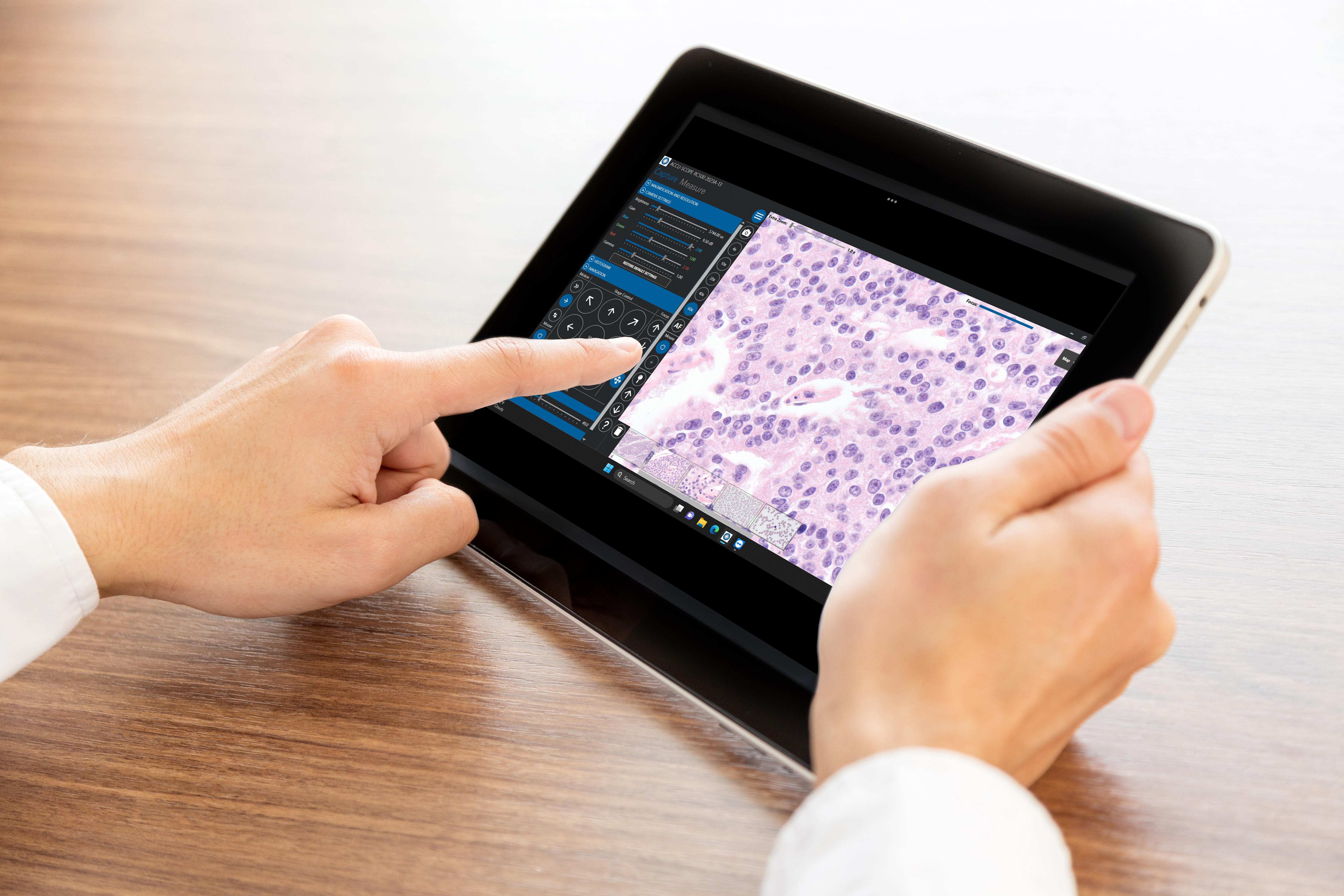 |
Real-Time TelepathologyReal-time telepathology systems stream LIVE digital video of glass slides to any remote location. Real-time systems use a motorized microscope, a high-resolution video camera and a streaming software to allow a remote consulting pathologist to control the microscope and evaluate a patient slide from any location at any time. |
 |
Rapid OFF-Site Evaluation (ROSE)Advances in digital imaging technology and development of high-speed internet have brought a shift in ROSE practice (rapid on-site evaluation) from the traditional in-person slide review to remote evaluation. Telecytology (TC) has increased the efficiency of pathologists by reducing travel time to procedure sites and eliminating long wait times between procedures. Today’s ROSE (rapid off-site evaluation) also provides the pathologist the flexibility to cover procedures occurring at several locations simultaneously. |
 |
Elevated Conferences, Training and CollaborationSimultaneous group viewing of images is necessary for a variety of instructional sessions and consultative meetings including conferences and tumor boards. The RC500 provides access to live images from the original glass slides. Both local and remote users have full control of the micrsocope -- including objectives, specimen navigation and image control -- to readily address the flow and needs of the discussion. |
 |
Doing More with LessNumerous studies show a trend in fewer pathologists (when adjusted for population and diagnosis levels) to do the important work that only they can do. In addition, the CMS reports reductions in the physician fee schedule (-1.25% for CY 2024). Pathologists and healthcare institutions are faced with the realization the they need to do more with less. Telepathology may be one solution to covering the case load and gaining access to pathologists and sub-specialists. The RC500 provides live telepathology of patient slides to anyone, anywhere at anytime. |
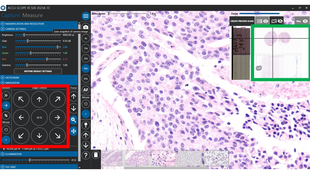 |
Fast, Easy NavigationRapid case reviews rely on fast and easy navigation of patient slides. The RC500 gives you multiple ways to drive.
Except for #4, all other navitation methods are available to both the remote reviewer and local user. |
Optional Accessories


 © 2022 ACCU-SCOPE® All rights reserved.