Microscopes are found in laboratories and science classrooms around the globe. With the advent of digital cameras, it is now even easier to upgrade a microscope to view the live image on a screen or monitor, capture images for documentation and analysis, and even stream live images with remote colleagues.
But not all cameras are alike, so how do you know which camera is best for you? Over the course of this multi-part series, we’ll try to explain the key specifications of cameras and why they matter. By the end, you should have a good idea of the best camera for your microscope type and application.
PART 1: Color or Monochrome
Let’s start with an easy question: Do you need a color or monochrome camera? If you are snapping photos of specimens with many different colors and you need to capture your specimens in all their colorful glory, then a color camera would be best.
But say your specimen is fluorescent, and it doesn’t appear very bright when viewed through the eyepieces. Even if you may want to view your specimen with different fluorescent channels (different excitation wavelengths and different emission wavelengths, therefore different colors), then a monochrome camera may be your best option. Let’s explore why. To create a color image, most color cameras use color filters in a distinctive pattern overlayered onto the pixels of the camera sensor. The Bayer mosaic mask is the most common color filter pattern (see Figure 1). The tiny filters are arranged in a 2x2 pattern with position 1 being blue, positions 2 and 3 are green, and position 4 is red. Firmware on the camera itself “fills in the gaps” so that every pixel is interpreted by a computer or monitor as having all three color components – red, blue and green.
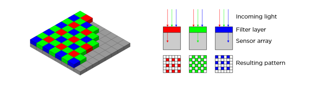
Source: https://en.wikipedia.org/wiki/Bayer_filter
By definition, a filter only allows some things to pass through it. In the case of a color camera, the pixels only “see” a little bit of the light that emanates from the sample. Monochrome cameras do not have these filters. For this reason, monochrome cameras are more sensitive than their color counterparts by allowing more light to reach the pixels on the sensor.
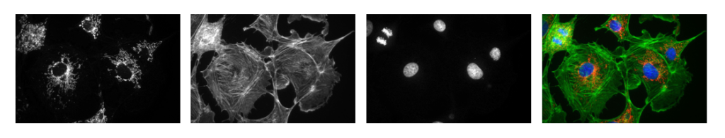
Just because monochrome cameras are “color blind” doesn’t mean that you can’t view the image in color. To make it more appealing to the human eye, most camera software allows the user to apply a color to the monochromatic image, often referred to as pseudocoloring. Most people will pseudocolor a monochrome image using a color similar to what they would see through the eyepieces (e.g., DAPI has peak emission at 461nm, so a blue of ~461nm is generally used). In Figure 2, the nuclei were stained with DAPI. The mitochondria were labeled with a red-emitting fluorophore, and the cytoskeleton was labeled with a green-fluorescing fluorophore. Each of those channels was pseudocolored according to the emission color of the fluorophore. Monochrome images, however, allow the user to choose any color to differentiate one stained structure in the sample from another.
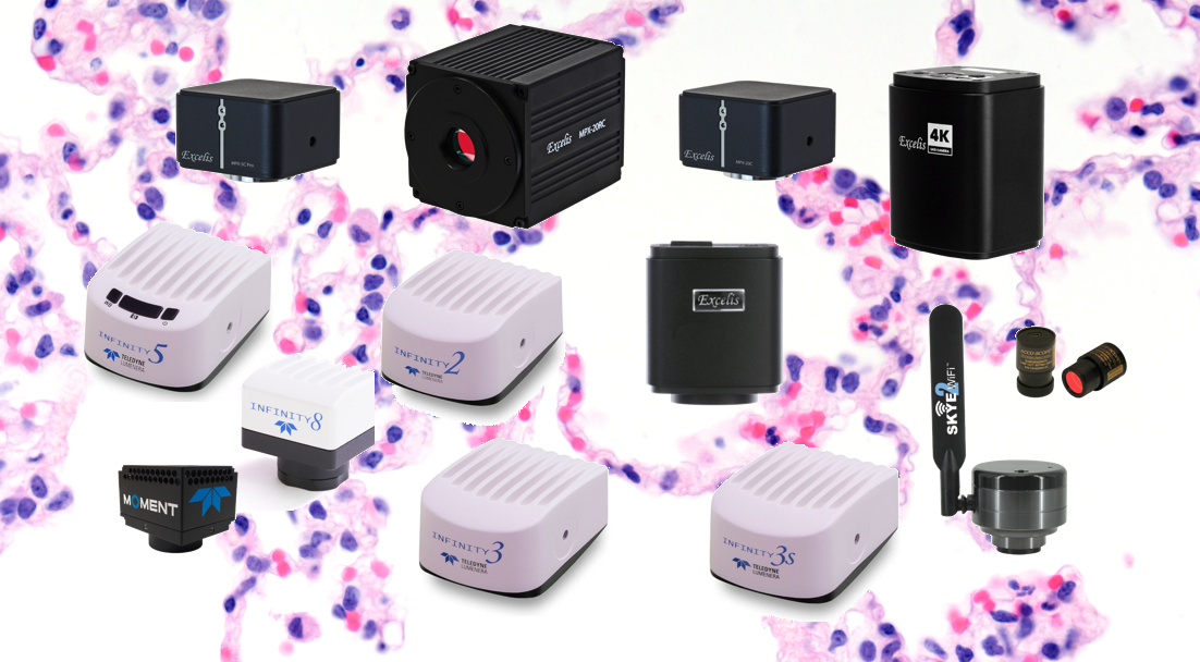
ACCU-SCOPE is proud to carry microscopy cameras from Teledyne Lumenera. Lumenera Infinity cameras are available in both color and monochrome versions. View the selection of Infinity cameras HERE. ACCU-SCOPE also offers color cameras for a wide variety of applications. View them HERE.
In the next part of our series, we’ll explore how your microscope configuration may impact your camera selection.
